 Movie
Movie Controller


+ Open data

- Basic information
| Entry | Database: PDB / ID: 1o1j | ||||||
|---|---|---|---|---|---|---|---|
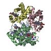
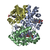
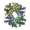
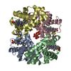
| Title | Deoxy hemoglobin (A-GLY-C:V1M,L29F,H58Q; B,D:V1M,L106W) | ||||||
 Components Components |
| ||||||
 Keywords Keywords | OXYGEN STORAGE/TRANSPORT / HEME / OXYGEN DELIVERY VEHICLE / BLOOD SUBSTITUTE / OXYGEN STORAGE-TRANSPORT COMPLEX | ||||||
| Function / homology |  Function and homology information Function and homology informationcellular oxidant detoxification / Heme assimilation / nitric oxide transport / hemoglobin alpha binding / hemoglobin binding / haptoglobin-hemoglobin complex / renal absorption / hemoglobin complex / oxygen transport / Scavenging of heme from plasma ...cellular oxidant detoxification / Heme assimilation / nitric oxide transport / hemoglobin alpha binding / hemoglobin binding / haptoglobin-hemoglobin complex / renal absorption / hemoglobin complex / oxygen transport / Scavenging of heme from plasma / erythrocyte development / endocytic vesicle lumen / blood vessel diameter maintenance / hydrogen peroxide catabolic process / oxygen carrier activity / carbon dioxide transport / response to hydrogen peroxide / Heme signaling / Erythrocytes take up oxygen and release carbon dioxide / Erythrocytes take up carbon dioxide and release oxygen / Cytoprotection by HMOX1 / oxygen binding / Late endosomal microautophagy / platelet aggregation / regulation of blood pressure / Chaperone Mediated Autophagy / positive regulation of nitric oxide biosynthetic process / tertiary granule lumen / Factors involved in megakaryocyte development and platelet production / blood microparticle / ficolin-1-rich granule lumen / iron ion binding / inflammatory response / heme binding / Neutrophil degranulation / : / extracellular exosome / extracellular region / membrane / metal ion binding / cytosol Similarity search - Function | ||||||
| Biological species |  Homo sapiens (human) Homo sapiens (human) | ||||||
| Method |  X-RAY DIFFRACTION / MOLECULAR REPLACEMENT / Resolution: 1.9 Å X-RAY DIFFRACTION / MOLECULAR REPLACEMENT / Resolution: 1.9 Å | ||||||
 Authors Authors | Brucker, E.A. | ||||||
 Citation Citation | #1: Journal: Acta Crystallogr.,Sect.D / Year: 2000Title: Genetically Crosslinked Hemoglobin: A Structural Study Authors: Brucker, E.A. #2: Journal: NAT.BIOTECHNOL. / Year: 1998Title: Rate of Reaction with Nitric Oxide Determines the Hypertensive Effect of Cell-Free Hemoglobin Authors: Doherty, D.H. / Doyle, M.P. / Curry, S.R. / Vali, R.J. / Fattor, T.J. / Olson, J.S. / Lemon, D.D. #3: Journal: Nature / Year: 1992Title: A Human Recombinant Hemoglobin Designed for Use as a Blood Substitute Authors: Looker, D. / Abbot-Brown, D. / Cozart, P. / Durfee, S. / Hoffman, S. / Mathews, A.J. / Miller-Roehrich, J. / Shoemaker, S. / Trimble, S. / Fermi, G. / Komiyama, N.H. / Nagai, K. / Stetler, G.L. | ||||||
| History |
| ||||||
| Remark 999 | SEQUENCE HUMAN HEMOGLOBIN HAS TWO ALPHA CHAINS WHICH ARE CALLED CHAINS A AND C IN OTHER PDB FILES. ...SEQUENCE HUMAN HEMOGLOBIN HAS TWO ALPHA CHAINS WHICH ARE CALLED CHAINS A AND C IN OTHER PDB FILES. IN THIS ENTRY THE TWO ALPHA CHAINS HAVE BEEN COVALENTLY JOINED TOGETHER BY ONE GLYCINE RESIDUE TO FORM ONE COVALENTLY LINKED POLYPEPTIDE CHAIN. |
- Structure visualization
| Structure viewer | Molecule: MolmilJmol/JSmol |
|---|
- Downloads & links
-Download
| PDBx/mmCIF format | 1o1j.cif.gz | 135.6 KB | Display | PDBx/mmCIF format |
|---|---|---|---|---|
| PDB format | pdb1o1j.ent.gz | 104.6 KB | Display | PDB format |
| PDBx/mmJSON format | 1o1j.json.gz | Tree view | PDBx/mmJSON format | |
| Others |  Other downloads Other downloads |
-Validation report
| Arichive directory | https://data.pdbj.org/pub/pdb/validation_reports/o1/1o1jftp://data.pdbj.org/pub/pdb/validation_reports/o1/1o1j | HTTPS FTP |
|---|
-Related structure data
| Related structure data |  1abyS S: Starting model for refinement |
|---|---|
| Similar structure data |


-Links
 PDBj
PDBj


- Assembly
| Deposited unit | 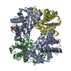
| ||||||||
|---|---|---|---|---|---|---|---|---|---|
| 1 |
| ||||||||
| Unit cell |
|
-Components
| #1: Protein | Mass: 30419.838 Da / Num. of mol.: 1 / Mutation: V1M, L29F, H58Q Source method: isolated from a genetically manipulated source Source: (gene. exp.) Homo sapiens (human) / Cell: RED BLOOD CELL / Production host:  | ||||
|---|---|---|---|---|---|
| #2: Protein | Mass: 15995.317 Da / Num. of mol.: 2 / Mutation: V1M, L106W Source method: isolated from a genetically manipulated source Source: (gene. exp.) Homo sapiens (human) / Cell: RED BLOOD CELL / Production host: #3: Chemical | ChemComp-HEM /   Mass: 616.487 Da / Num. of mol.: 4 / Source method: obtained synthetically / Formula: C34H32FeN4O4 Mass: 616.487 Da / Num. of mol.: 4 / Source method: obtained synthetically / Formula: C34H32FeN4O4#4: Water | ChemComp-HOH / |  Mass: 18.015 Da / Num. of mol.: 370 / Source method: isolated from a natural source / Formula: H2O Mass: 18.015 Da / Num. of mol.: 370 / Source method: isolated from a natural source / Formula: H2O |
-Experimental details
-Experiment
| Experiment | Method: X-RAY DIFFRACTION / Number of used crystals: 1 |
|---|
- Sample preparation
| Crystal | Density Matthews: 1.84 Å3/Da / Density % sol: 33.2 % |
|---|---|
| Crystal grow | pH: 6.5 / Details: pH 6.50 |
-Data collection
| Diffraction | Mean temperature: 295 K |
|---|---|
| Diffraction source | Source: ROTATING ANODE / Type: SIEMENS / Wavelength: 1.5418 |
| Detector | Type: RIGAKU RAXIS IIC / Detector: IMAGE PLATE / Date: Apr 15, 1998 / Details: COLLIMATOR |
| Radiation | Monochromator: GRAPHITE / Protocol: SINGLE WAVELENGTH / Monochromatic (M) / Laue (L): M / Scattering type: x-ray |
| Radiation wavelength | Wavelength: 1.5418 Å / Relative weight: 1 |
| Reflection | Resolution: 1.9→30 Å / Num. obs: 39493 / % possible obs: 90.7 % / Redundancy: 3.54 % / Rmerge(I) obs: 0.055 / Net I/σ(I): 27.3 |
| Reflection shell | Resolution: 1.9→2 Å / Redundancy: 3.06 % / Rmerge(I) obs: 0.304 / Mean I/σ(I) obs: 7.15 / % possible all: 79.2 |
- Processing
| Software |
| |||||||||||||||||||||||||||||||||
|---|---|---|---|---|---|---|---|---|---|---|---|---|---|---|---|---|---|---|---|---|---|---|---|---|---|---|---|---|---|---|---|---|---|---|
| Refinement | Method to determine structure: MOLECULAR REPLACEMENT Starting model: PDB ENTRY 1ABY Resolution: 1.9→8 Å / Num. parameters: 19795 / Num. restraintsaints: 23143 / Cross valid method: THROUGHOUT / σ(F): 0 / Stereochemistry target values: ENGH AND HUBER
| |||||||||||||||||||||||||||||||||
| Solvent computation | Solvent model: MOEWS & KRETSINGER, J.MOL.BIOL. 91(1973) 201-228 | |||||||||||||||||||||||||||||||||
| Refine analyze | Num. disordered residues: 0 / Occupancy sum hydrogen: 0 / Occupancy sum non hydrogen: 4941 | |||||||||||||||||||||||||||||||||
| Refinement step | Cycle: LAST / Resolution: 1.9→8 Å
| |||||||||||||||||||||||||||||||||
| Refine LS restraints |
|
