 Movie
Movie Controller


+ Open data

- Basic information
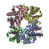
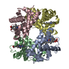
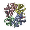
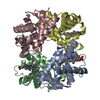
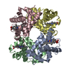
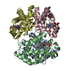
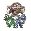
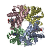
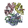
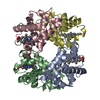
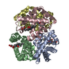
| Entry | Database: PDB / ID: 2hbs | ||||||
|---|---|---|---|---|---|---|---|
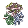
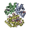
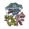
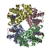
| Title | THE HIGH RESOLUTION CRYSTAL STRUCTURE OF DEOXYHEMOGLOBIN S | ||||||
 Components Components |
| ||||||
 Keywords Keywords | OXYGEN TRANSPORT / HEMOGLOBIN | ||||||
| Function / homology |  Function and homology information Function and homology informationcellular oxidant detoxification / Heme assimilation / nitric oxide transport / hemoglobin alpha binding / hemoglobin binding / haptoglobin-hemoglobin complex / renal absorption / hemoglobin complex / oxygen transport / Scavenging of heme from plasma ...cellular oxidant detoxification / Heme assimilation / nitric oxide transport / hemoglobin alpha binding / hemoglobin binding / haptoglobin-hemoglobin complex / renal absorption / hemoglobin complex / oxygen transport / Scavenging of heme from plasma / erythrocyte development / endocytic vesicle lumen / blood vessel diameter maintenance / hydrogen peroxide catabolic process / oxygen carrier activity / carbon dioxide transport / response to hydrogen peroxide / Heme signaling / Erythrocytes take up oxygen and release carbon dioxide / Erythrocytes take up carbon dioxide and release oxygen / Cytoprotection by HMOX1 / oxygen binding / Late endosomal microautophagy / platelet aggregation / regulation of blood pressure / Chaperone Mediated Autophagy / positive regulation of nitric oxide biosynthetic process / tertiary granule lumen / Factors involved in megakaryocyte development and platelet production / blood microparticle / ficolin-1-rich granule lumen / iron ion binding / inflammatory response / heme binding / Neutrophil degranulation / : / extracellular exosome / extracellular region / membrane / metal ion binding / cytosol Similarity search - Function | ||||||
| Biological species |  Homo sapiens (human) Homo sapiens (human) | ||||||
| Method |  X-RAY DIFFRACTION / Resolution: 2.05 Å X-RAY DIFFRACTION / Resolution: 2.05 Å | ||||||
 Authors Authors | Harrington, D.J. / Adachi, K. / Royer Junior, W.E. | ||||||
 Citation Citation | Journal: J.Mol.Biol. / Year: 1997 Title: The high resolution crystal structure of deoxyhemoglobin S. Authors: Harrington, D.J. / Adachi, K. / Royer Jr., W.E. #1: Journal: J.Biol.Chem. / Year: 1985Title: Refined Crystal Structure of Deoxyhemoglobin S. II. Molecular Interactions in the Crystal Authors: Padlan, E.A. / Love, W.E. #2: Journal: J.Biol.Chem. / Year: 1985Title: Refined Crystal Structure of Deoxyhemoglobin S. I. Restrained Least-Squares Refinement at 3.0-A Resolution Authors: Padlan, E.A. / Love, W.E. #3: Journal: Inserm Symp. / Year: 1978Title: Intermolecular Interactions in Crystals of Human Deoxy Hemoglobin A, C, F and S Authors: Love, W.E. / Fitzgerald, P.M.D. / Hanson, J.C. / Royer Junior, W.E. #4: Journal: Proceedings of the Symposium on Molecular and Cellular Aspects of Sickle Cell Disease, Dallas, Texas, December 10, 1975 (in: Dhew Pub (Nih) (Us), No.76-1007)Year: 1976 Title: Crystal Structure of Sickle-Cell Deoxyhemoglobin Authors: Wishner, B.C. / Hanson, J.C. / Ringle, W.M. / Love, W.E. #5: Journal: Proceedings of the First National Symposium on Sickle Cell Disease, Washington, D.C., June 27-29, 1974 (in: Dhew Pub (Nih) (Us), No.75-723)Year: 1975 Title: Crystals of Deoxy Sickle Cell Hemoglobin Authors: Wishner, B.C. / Love, W.E. #6: Journal: J.Mol.Biol. / Year: 1975Title: Crystal Structure of Sickle-Cell Deoxyhemoglobin at 5 A Resolution Authors: Wishner, B.C. / Ward, K.B. / Lattman, E.E. / Love, W.E. | ||||||
| History |
|
- Structure visualization
| Structure viewer | Molecule: MolmilJmol/JSmol |
|---|
- Downloads & links
-Download
| PDBx/mmCIF format | 2hbs.cif.gz | 242 KB | Display | PDBx/mmCIF format |
|---|---|---|---|---|
| PDB format | pdb2hbs.ent.gz | 198.1 KB | Display | PDB format |
| PDBx/mmJSON format | 2hbs.json.gz | Tree view | PDBx/mmJSON format | |
| Others |  Other downloads Other downloads |
-Validation report
| Arichive directory | https://data.pdbj.org/pub/pdb/validation_reports/hb/2hbsftp://data.pdbj.org/pub/pdb/validation_reports/hb/2hbs | HTTPS FTP |
|---|
-Related structure data
| Similar structure data |
|---|


-Links
 PDBj
PDBj


- Assembly
| Deposited unit | 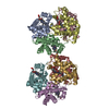
| ||||||||
|---|---|---|---|---|---|---|---|---|---|
| 1 | 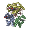
| ||||||||
| 2 | 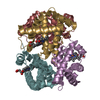
| ||||||||
| Unit cell |
| ||||||||
| Noncrystallographic symmetry (NCS) | NCS oper: (Code: given Matrix: (0.9997, -0.0211, 0.01274), Vector: |
-Components
| #1: Protein | Mass: 15150.353 Da / Num. of mol.: 4 / Source method: isolated from a natural source / Source: (natural) Homo sapiens (human) / Cell: RED BLOOD CELLS / Cellular location: CYTOPLASM / Organ: BLOOD / Tissue: BLOOD / References: UniProt: P69905#2: Protein | Mass: 15860.216 Da / Num. of mol.: 4 / Mutation: E6V VARIANT / Source method: isolated from a natural source / Source: (natural) Homo sapiens (human) / Cell: RED BLOOD CELLS / Cellular location: CYTOPLASM / Organ: BLOOD / Tissue: BLOOD / References: UniProt: P68871#3: Chemical | ChemComp-HEM /   Mass: 616.487 Da / Num. of mol.: 8 / Source method: obtained synthetically / Formula: C34H32FeN4O4 Mass: 616.487 Da / Num. of mol.: 8 / Source method: obtained synthetically / Formula: C34H32FeN4O4#4: Water | ChemComp-HOH / |  Mass: 18.015 Da / Num. of mol.: 573 / Source method: isolated from a natural source / Formula: H2O Mass: 18.015 Da / Num. of mol.: 573 / Source method: isolated from a natural source / Formula: H2O |
|---|
-Experimental details
-Experiment
| Experiment | Method: X-RAY DIFFRACTION / Number of used crystals: 3 |
|---|
- Sample preparation
| Crystal | Density Matthews: 2.51 Å3/Da / Density % sol: 50.91 % Description: HIGH RESOLUTION DATA USED TO REFINE 1HBS STRUCTURE | |||||||||||||||
|---|---|---|---|---|---|---|---|---|---|---|---|---|---|---|---|---|
| Crystal grow | pH: 4 Details: HBS WAS SUSPENDED IN 30 MM PHOSPHATE BUFFER, PH 7.0, DEOXYGENATED AND CONCENTRATED TO 120 MG/ML. PROTEIN WAS CRYSTALLIZED IN AN ANAEROBIC CHAMBER IN TUBE CONTAINING 10 UL PROTEIN SOLUTION, 5 ...Details: HBS WAS SUSPENDED IN 30 MM PHOSPHATE BUFFER, PH 7.0, DEOXYGENATED AND CONCENTRATED TO 120 MG/ML. PROTEIN WAS CRYSTALLIZED IN AN ANAEROBIC CHAMBER IN TUBE CONTAINING 10 UL PROTEIN SOLUTION, 5 UL CITRATE BUFFER (PH 4.0), AND 4UL OF 33% PEG 8K, crystallized in anaerobic chamber | |||||||||||||||
| Crystal grow | *PLUS Method: unknownDetails: refer to Wishner, B.C., (1976) Proceedings of the Symposium on Molecular and Cellular Aspects of Sickle Cell Disease, pp. 1-31. | |||||||||||||||
| Components of the solutions | *PLUS
|
-Data collection
| Diffraction | Mean temperature: 295 K |
|---|---|
| Diffraction source | Source: ROTATING ANODE / Type: RIGAKU RUH2R / Wavelength: 1.5418 |
| Detector | Type: RIGAKU / Detector: IMAGE PLATE / Date: Mar 9, 1995 / Details: COLLIMATOR |
| Radiation | Monochromator: GRAPHITE(002) / Monochromatic (M) / Laue (L): M / Scattering type: x-ray |
| Radiation wavelength | Wavelength: 1.5418 Å / Relative weight: 1 |
| Reflection | Resolution: 2.05→100 Å / Num. obs: 72955 / % possible obs: 95 % / Observed criterion σ(I): 2 / Redundancy: 3.6 % / Biso Wilson estimate: 16.1 Å2 / Rsym value: 0.093 |
| Reflection shell | Resolution: 2.05→2.18 Å / Rsym value: 0.371 / % possible all: 86.1 |
| Reflection | *PLUS Rmerge(I) obs: 0.093 |
| Reflection shell | *PLUS % possible obs: 86.1 % / Rmerge(I) obs: 0.371 |
- Processing
| Software |
| ||||||||||||||||||||||||||||||||||||||||||||||||||||||||||||||||||||||||||||||||
|---|---|---|---|---|---|---|---|---|---|---|---|---|---|---|---|---|---|---|---|---|---|---|---|---|---|---|---|---|---|---|---|---|---|---|---|---|---|---|---|---|---|---|---|---|---|---|---|---|---|---|---|---|---|---|---|---|---|---|---|---|---|---|---|---|---|---|---|---|---|---|---|---|---|---|---|---|---|---|---|---|---|
| Refinement | Resolution: 2.05→10 Å / Rfactor Rfree error: 0.003 / Data cutoff high absF: 100000 / Data cutoff low absF: 0.1 / Isotropic thermal model: RESTRAINED / Cross valid method: THROUGHOUT / σ(F): 2 / Details: BULK SOLVENT MODEL USED
| ||||||||||||||||||||||||||||||||||||||||||||||||||||||||||||||||||||||||||||||||
| Displacement parameters | Biso mean: 20.5 Å2 | ||||||||||||||||||||||||||||||||||||||||||||||||||||||||||||||||||||||||||||||||
| Refine analyze | Luzzati coordinate error obs: 0.22 Å | ||||||||||||||||||||||||||||||||||||||||||||||||||||||||||||||||||||||||||||||||
| Refinement step | Cycle: LAST / Resolution: 2.05→10 Å
| ||||||||||||||||||||||||||||||||||||||||||||||||||||||||||||||||||||||||||||||||
| Refine LS restraints |
| ||||||||||||||||||||||||||||||||||||||||||||||||||||||||||||||||||||||||||||||||
| LS refinement shell | Resolution: 2.05→2.14 Å / Rfactor Rfree error: 0.012 / Total num. of bins used: 8
| ||||||||||||||||||||||||||||||||||||||||||||||||||||||||||||||||||||||||||||||||
| Xplor file |
| ||||||||||||||||||||||||||||||||||||||||||||||||||||||||||||||||||||||||||||||||
| Software | *PLUS Name: X-PLOR / Version: 3.1 / Classification: refinement | ||||||||||||||||||||||||||||||||||||||||||||||||||||||||||||||||||||||||||||||||
| Refine LS restraints | *PLUS
|
