 Movie
Movie Controller


+ Open data

- Basic information
| Entry | Database: PDB / ID: 1o1m | ||||||
|---|---|---|---|---|---|---|---|
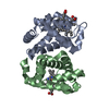
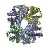
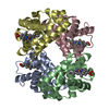
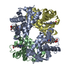
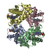
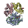
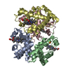
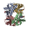
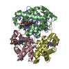
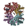
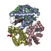
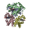
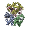
| Title | Deoxy hemoglobin (A-GLYGLYGLY-C:V1M,L29F,H58Q B,D:V1M,V67W) | ||||||
 Components Components |
| ||||||
 Keywords Keywords | OXYGEN STORAGE/TRANSPORT / HEME / OXYGEN DELIVERY VEHICLE / BLOOD SUBSTITUTE / OXYGEN STORAGE-TRANSPORT COMPLEX | ||||||
| Function / homology |  Function and homology information Function and homology informationcellular oxidant detoxification / Heme assimilation / nitric oxide transport / hemoglobin alpha binding / hemoglobin binding / haptoglobin-hemoglobin complex / renal absorption / hemoglobin complex / oxygen transport / Scavenging of heme from plasma ...cellular oxidant detoxification / Heme assimilation / nitric oxide transport / hemoglobin alpha binding / hemoglobin binding / haptoglobin-hemoglobin complex / renal absorption / hemoglobin complex / oxygen transport / Scavenging of heme from plasma / erythrocyte development / endocytic vesicle lumen / blood vessel diameter maintenance / hydrogen peroxide catabolic process / oxygen carrier activity / carbon dioxide transport / response to hydrogen peroxide / Heme signaling / Erythrocytes take up oxygen and release carbon dioxide / Erythrocytes take up carbon dioxide and release oxygen / Cytoprotection by HMOX1 / oxygen binding / Late endosomal microautophagy / platelet aggregation / regulation of blood pressure / Chaperone Mediated Autophagy / positive regulation of nitric oxide biosynthetic process / tertiary granule lumen / Factors involved in megakaryocyte development and platelet production / blood microparticle / ficolin-1-rich granule lumen / iron ion binding / inflammatory response / heme binding / Neutrophil degranulation / : / extracellular exosome / extracellular region / membrane / metal ion binding / cytosol Similarity search - Function | ||||||
| Biological species |  Homo sapiens (human) Homo sapiens (human) | ||||||
| Method |  X-RAY DIFFRACTION / MOLECULAR REPLACEMENT / Resolution: 1.85 Å X-RAY DIFFRACTION / MOLECULAR REPLACEMENT / Resolution: 1.85 Å | ||||||
 Authors Authors | Brucker, E.A. | ||||||
 Citation Citation | #1: Journal: Acta Crystallogr.,Sect.D / Year: 2000Title: Genetically Crosslinked Hemoglobin: A Structural Study Authors: Brucker, E.A. #2: Journal: NAT.BIOTECHNOL. / Year: 1998Title: Rate of Reaction with Nitric Oxide Determines the Hypertensive Effect of Cell-Free Hemoglobin Authors: Doherty, D.H. / Doyle, M.P. / Curry, S.R. / Vali, R.J. / Fattor, T.J. / Olson, J.S. / Lemon, D.D. #3: Journal: Nature / Year: 1992Title: A Human Recombinant Hemoglobin Designed for Use as a Blood Substitute Authors: Looker, D. / Abbot-Brown, D. / Cozart, P. / Durfee, S. / Hoffman, S. / Mathews, A.J. / Miller-Roehrich, J. / Shoemaker, S. / Trimble, S. / Fermi, G. / Komiyama, N.H. / Nagai, K. / Stetler, G.L. | ||||||
| History |
| ||||||
| Remark 999 | SEQUENCE HUMAN HEMOGLOBIN HAS TWO ALPHA CHAINS WHICH ARE CALLED CHAINS A AND C IN OTHER PDB FILES. ...SEQUENCE HUMAN HEMOGLOBIN HAS TWO ALPHA CHAINS WHICH ARE CALLED CHAINS A AND C IN OTHER PDB FILES. IN THIS ENTRY THE C-TERMINUS OF THE ALPHA-1 CHAIN AND THE N-TERMINUS OF THE ALPHA-2 CHAIN ARE GENETICALLY LINKED BY THREE GLYCINE RESIDUES TO FORM ONE COVALENTLY LINKED POLYPEPTIDE CHAIN. THESE THREE RESIDUES ARE HIGHLY DISORDERED, I.E. NOT DEFINED IN ELECTRON DENSITY. ALSO, THE INITIAL RESIDUE OF THE ALPHA-2 CHAIN, JUST AFTER THE GLYCINE LINKER, IS THE NATIVE VALINE, NOT A METHIONINE. THEREFORE, THE THREE GLYCINE LINKER IS NOT INCLUDED IN THESE COORDINATES, AND THE INITIAL RESIDUE OF THE ALPHA CHAIN(S) IS MODELED AS AN ALANINE. |
- Structure visualization
| Structure viewer | Molecule: MolmilJmol/JSmol |
|---|
- Downloads & links
-Download
| PDBx/mmCIF format | 1o1m.cif.gz | 135 KB | Display | PDBx/mmCIF format |
|---|---|---|---|---|
| PDB format | pdb1o1m.ent.gz | 104.5 KB | Display | PDB format |
| PDBx/mmJSON format | 1o1m.json.gz | Tree view | PDBx/mmJSON format | |
| Others |  Other downloads Other downloads |
-Validation report
| Arichive directory | https://data.pdbj.org/pub/pdb/validation_reports/o1/1o1mftp://data.pdbj.org/pub/pdb/validation_reports/o1/1o1m | HTTPS FTP |
|---|
-Related structure data
| Related structure data |  1c7cS S: Starting model for refinement |
|---|---|
| Similar structure data |


-Links
 PDBj
PDBj


- Assembly
| Deposited unit | 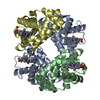
| ||||||||
|---|---|---|---|---|---|---|---|---|---|
| 1 |
| ||||||||
| Unit cell |
|
-Components
| #1: Protein | Mass: 30533.941 Da / Num. of mol.: 1 / Mutation: V1M, L29F, H58Q Source method: isolated from a genetically manipulated source Source: (gene. exp.) Homo sapiens (human) / Cell: RED BLOOD CELL / Production host:  | ||||
|---|---|---|---|---|---|
| #2: Protein | Mass: 16009.343 Da / Num. of mol.: 2 / Mutation: V1M, V67W Source method: isolated from a genetically manipulated source Source: (gene. exp.) Homo sapiens (human) / Cell: RED BLOOD CELL / Production host: #3: Chemical | ChemComp-HEM /   Mass: 616.487 Da / Num. of mol.: 4 / Source method: obtained synthetically / Formula: C34H32FeN4O4 Mass: 616.487 Da / Num. of mol.: 4 / Source method: obtained synthetically / Formula: C34H32FeN4O4#4: Water | ChemComp-HOH / |  Mass: 18.015 Da / Num. of mol.: 339 / Source method: isolated from a natural source / Formula: H2O Mass: 18.015 Da / Num. of mol.: 339 / Source method: isolated from a natural source / Formula: H2O |
-Experimental details
-Experiment
| Experiment | Method: X-RAY DIFFRACTION / Number of used crystals: 1 |
|---|
- Sample preparation
| Crystal | Density Matthews: 1.85 Å3/Da / Density % sol: 33.5 % |
|---|---|
| Crystal grow | pH: 6.5 / Details: pH 6.50 |
-Data collection
| Diffraction | Mean temperature: 295 K |
|---|---|
| Diffraction source | Source: ROTATING ANODE / Type: SIEMENS / Wavelength: 1.5418 |
| Detector | Type: RIGAKU RAXIS IIC / Detector: IMAGE PLATE / Date: Aug 15, 1998 / Details: COLLIMATOR |
| Radiation | Monochromator: GRAPHITE / Protocol: SINGLE WAVELENGTH / Monochromatic (M) / Laue (L): M / Scattering type: x-ray |
| Radiation wavelength | Wavelength: 1.5418 Å / Relative weight: 1 |
| Reflection | Resolution: 1.85→30 Å / Num. obs: 43765 / % possible obs: 92.7 % / Redundancy: 2.51 % / Rmerge(I) obs: 0.049 / Net I/σ(I): 24.7 |
| Reflection shell | Resolution: 1.85→1.95 Å / Redundancy: 2.3 % / Rmerge(I) obs: 0.277 / Mean I/σ(I) obs: 5.01 / % possible all: 93.7 |
- Processing
| Software |
| |||||||||||||||||||||||||||||||||
|---|---|---|---|---|---|---|---|---|---|---|---|---|---|---|---|---|---|---|---|---|---|---|---|---|---|---|---|---|---|---|---|---|---|---|
| Refinement | Method to determine structure: MOLECULAR REPLACEMENT Starting model: pdb entry 1C7C Resolution: 1.85→8 Å / Num. parameters: 19681 / Num. restraintsaints: 23153 / Cross valid method: THROUGHOUT / σ(F): 0 / Stereochemistry target values: ENGH AND HUBER
| |||||||||||||||||||||||||||||||||
| Solvent computation | Solvent model: MOEWS & KRETSINGER, J.MOL.BIOL. 91(1973) 201-228 | |||||||||||||||||||||||||||||||||
| Refine analyze | Num. disordered residues: 2 / Occupancy sum hydrogen: 0 / Occupancy sum non hydrogen: 4895 | |||||||||||||||||||||||||||||||||
| Refinement step | Cycle: LAST / Resolution: 1.85→8 Å
| |||||||||||||||||||||||||||||||||
| Refine LS restraints |
|
