 Movie
Movie Controller


+ Open data

- Basic information
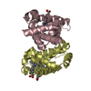
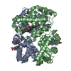
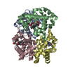
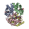
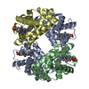
| Entry | Database: PDB / ID: 1a9w | ||||||
|---|---|---|---|---|---|---|---|
| Title | HUMAN EMBRYONIC GOWER II CARBONMONOXY HEMOGLOBIN | ||||||
 Components Components |
| ||||||
 Keywords Keywords | OXYGEN TRANSPORT | ||||||
| Function / homology |  Function and homology information Function and homology informationcellular oxidant detoxification / Heme assimilation / nitric oxide transport / hemoglobin alpha binding / haptoglobin-hemoglobin complex / hemoglobin complex / oxygen transport / Scavenging of heme from plasma / erythrocyte development / endocytic vesicle lumen ...cellular oxidant detoxification / Heme assimilation / nitric oxide transport / hemoglobin alpha binding / haptoglobin-hemoglobin complex / hemoglobin complex / oxygen transport / Scavenging of heme from plasma / erythrocyte development / endocytic vesicle lumen / hydrogen peroxide catabolic process / oxygen carrier activity / carbon dioxide transport / response to hydrogen peroxide / Heme signaling / Erythrocytes take up oxygen and release carbon dioxide / Erythrocytes take up carbon dioxide and release oxygen / Cytoprotection by HMOX1 / oxygen binding / Factors involved in megakaryocyte development and platelet production / blood microparticle / iron ion binding / inflammatory response / heme binding / : / extracellular exosome / extracellular region / membrane / metal ion binding / cytosol Similarity search - Function | ||||||
| Biological species |  Homo sapiens (human) Homo sapiens (human) | ||||||
| Method |  X-RAY DIFFRACTION / MOLECULAR REPLACEMENT / Resolution: 2.9 Å X-RAY DIFFRACTION / MOLECULAR REPLACEMENT / Resolution: 2.9 Å | ||||||
 Authors Authors | Sutherland-Smith, A.J. / Baker, H.M. / Hofmann, O.M. / Brittain, T. / Baker, E.N. | ||||||
 Citation Citation | Journal: J.Mol.Biol. / Year: 1998 Title: Crystal structure of a human embryonic haemoglobin: the carbonmonoxy form of gower II (alpha2 epsilon2) haemoglobin at 2.9 A resolution. Authors: Sutherland-Smith, A.J. / Baker, H.M. / Hofmann, O.M. / Brittain, T. / Baker, E.N. | ||||||
| History |
|
- Structure visualization
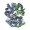
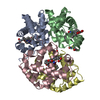
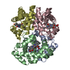
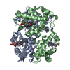
| Structure viewer | Molecule: MolmilJmol/JSmol |
|---|
- Downloads & links
-Download
| PDBx/mmCIF format | 1a9w.cif.gz | 112.6 KB | Display | PDBx/mmCIF format |
|---|---|---|---|---|
| PDB format | pdb1a9w.ent.gz | 88.9 KB | Display | PDB format |
| PDBx/mmJSON format | 1a9w.json.gz | Tree view | PDBx/mmJSON format | |
| Others |  Other downloads Other downloads |
-Validation report
| Arichive directory | https://data.pdbj.org/pub/pdb/validation_reports/a9/1a9wftp://data.pdbj.org/pub/pdb/validation_reports/a9/1a9w | HTTPS FTP |
|---|
-Related structure data
| Related structure data | 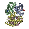 1bbbS S: Starting model for refinement |
|---|---|
| Similar structure data |

-Links
 PDBj
PDBj


- Assembly
| Deposited unit | 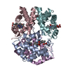
| |||||||||||||||||||||
|---|---|---|---|---|---|---|---|---|---|---|---|---|---|---|---|---|---|---|---|---|---|---|
| 1 |
| |||||||||||||||||||||
| Unit cell |
| |||||||||||||||||||||
| Noncrystallographic symmetry (NCS) | NCS domain:
NCS oper:
|
-Components
| #1: Protein | Mass: 15150.353 Da / Num. of mol.: 2 Source method: isolated from a genetically manipulated source Details: HUMAN GOWER II EMBRYONIC HEMOGLOBIN / Source: (gene. exp.) Homo sapiens (human) / Plasmid: PRMAE389 / Production host:  #2: Protein | Mass: 16094.614 Da / Num. of mol.: 2 Source method: isolated from a genetically manipulated source Details: HUMAN GOWER II EMBRYONIC HEMOGLOBIN / Source: (gene. exp.) Homo sapiens (human) / Plasmid: PRMAE389 / Production host: #3: Chemical | ChemComp-HEM /   Mass: 616.487 Da / Num. of mol.: 4 / Source method: obtained synthetically / Formula: C34H32FeN4O4 Mass: 616.487 Da / Num. of mol.: 4 / Source method: obtained synthetically / Formula: C34H32FeN4O4#4: Chemical | ChemComp-CMO /   Mass: 28.010 Da / Num. of mol.: 4 / Source method: obtained synthetically / Formula: CO Mass: 28.010 Da / Num. of mol.: 4 / Source method: obtained synthetically / Formula: CO |
|---|
-Experimental details
-Experiment
| Experiment | Method: X-RAY DIFFRACTION / Number of used crystals: 1 |
|---|
- Sample preparation
| Crystal | Density Matthews: 2.47 Å3/Da / Density % sol: 51 % | ||||||||||||||||||||||||||||||
|---|---|---|---|---|---|---|---|---|---|---|---|---|---|---|---|---|---|---|---|---|---|---|---|---|---|---|---|---|---|---|---|
| Crystal grow | Method: microseeding, macroseeding used to increase crystal size pH: 8.5 Details: PROTEIN WAS CRYSTALLIZED FROM 21% MME-PEG 5000, 0.2M TAPS/ KOH, PH 8.5, 2MM DITHIONITE. THEN MICROSEEDING AND MACROSEEDING USED TO INCREASE CRYSTAL SIZE, microseeding and macroseeding used ...Details: PROTEIN WAS CRYSTALLIZED FROM 21% MME-PEG 5000, 0.2M TAPS/ KOH, PH 8.5, 2MM DITHIONITE. THEN MICROSEEDING AND MACROSEEDING USED TO INCREASE CRYSTAL SIZE, microseeding and macroseeding used to increase crystal size | ||||||||||||||||||||||||||||||
| Crystal grow | *PLUS Method: vapor diffusion, hanging dropDetails: drop solution was mixed with an equal volume of reservoir solution | ||||||||||||||||||||||||||||||
| Components of the solutions | *PLUS
|
-Data collection
| Diffraction | Mean temperature: 287 K |
|---|---|
| Diffraction source | Source: ROTATING ANODE / Type: RIGAKU RUH2R / Wavelength: 1.5418 |
| Detector | Type: RIGAKU RAXIS IIC / Detector: IMAGE PLATE / Date: Oct 1, 1994 / Details: 0.3 MM COLLIMATOR |
| Radiation | Monochromator: GRAPHITE(002) / Monochromatic (M) / Laue (L): M / Scattering type: x-ray |
| Radiation wavelength | Wavelength: 1.5418 Å / Relative weight: 1 |
| Reflection | Resolution: 2.9→40 Å / Num. obs: 14645 / % possible obs: 95.9 % / Observed criterion σ(I): 1.9 / Redundancy: 5.9 % / Biso Wilson estimate: 61.7 Å2 / Rmerge(I) obs: 0.091 / Rsym value: 0.091 / Net I/σ(I): 7.1 |
| Reflection shell | Resolution: 2.9→2.97 Å / Redundancy: 4.5 % / Rmerge(I) obs: 0.37 / Mean I/σ(I) obs: 1.9 / Rsym value: 0.37 / % possible all: 76.3 |
| Reflection | *PLUS Num. obs: 14597 / Num. measured all: 118919 |
| Reflection shell | *PLUS % possible obs: 72.6 % / Rmerge(I) obs: 0.417 |
- Processing
| Software |
| ||||||||||||||||||||||||||||||||||||||||||||||||||||||||||||
|---|---|---|---|---|---|---|---|---|---|---|---|---|---|---|---|---|---|---|---|---|---|---|---|---|---|---|---|---|---|---|---|---|---|---|---|---|---|---|---|---|---|---|---|---|---|---|---|---|---|---|---|---|---|---|---|---|---|---|---|---|---|
| Refinement | Method to determine structure: MOLECULAR REPLACEMENT Starting model: PDB ENTRY 1BBB Resolution: 2.9→40 Å / Rfactor Rfree error: 0.006 / Data cutoff high absF: 1000000 / Data cutoff low absF: 0.001 Isotropic thermal model: ONE B FOR MAIN CHAIN ATOM B FOR SIDE CHAIN ATOMS Cross valid method: THROUGHOUT / σ(F): 0 / Details: TNT-5E ALSO USED
| ||||||||||||||||||||||||||||||||||||||||||||||||||||||||||||
| Displacement parameters | Biso mean: 50.3 Å2
| ||||||||||||||||||||||||||||||||||||||||||||||||||||||||||||
| Refine analyze | Luzzati sigma a obs: 0.35 Å | ||||||||||||||||||||||||||||||||||||||||||||||||||||||||||||
| Refinement step | Cycle: LAST / Resolution: 2.9→40 Å
| ||||||||||||||||||||||||||||||||||||||||||||||||||||||||||||
| Refine LS restraints |
| ||||||||||||||||||||||||||||||||||||||||||||||||||||||||||||
| Refine LS restraints NCS |
| ||||||||||||||||||||||||||||||||||||||||||||||||||||||||||||
| LS refinement shell | Resolution: 2.9→3.03 Å / Rfactor Rfree error: 0.03 / Total num. of bins used: 8
| ||||||||||||||||||||||||||||||||||||||||||||||||||||||||||||
| Xplor file |
| ||||||||||||||||||||||||||||||||||||||||||||||||||||||||||||
| Software | *PLUS Name: X-PLOR / Version: 3.851 / Classification: refinement | ||||||||||||||||||||||||||||||||||||||||||||||||||||||||||||
| Refinement | *PLUS Rfactor obs: 0.186 / Rfactor Rfree: 0.235 | ||||||||||||||||||||||||||||||||||||||||||||||||||||||||||||
| Solvent computation | *PLUS | ||||||||||||||||||||||||||||||||||||||||||||||||||||||||||||
| Displacement parameters | *PLUS | ||||||||||||||||||||||||||||||||||||||||||||||||||||||||||||
| Refine LS restraints | *PLUS
| ||||||||||||||||||||||||||||||||||||||||||||||||||||||||||||
| LS refinement shell | *PLUS Lowest resolution: 2.97 Å / Rfactor Rfree: 0.366 / Rfactor obs: 0.301 |
