 Movie
Movie Controller

[English] 日本語
 Yorodumi
- PDB-1bbb: A THIRD QUATERNARY STRUCTURE OF HUMAN HEMOGLOBIN A AT 1.7-ANGSTRO... -
+ Open data

- Basic information
| Entry | Database: PDB / ID: 1bbb | |||||||||
|---|---|---|---|---|---|---|---|---|---|---|
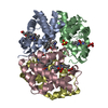
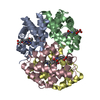
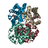
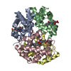
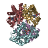
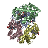
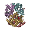
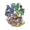
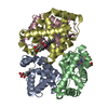
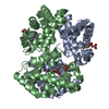
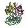
| Title | A THIRD QUATERNARY STRUCTURE OF HUMAN HEMOGLOBIN A AT 1.7-ANGSTROMS RESOLUTION | |||||||||
 Components Components |
| |||||||||
 Keywords Keywords | OXYGEN TRANSPORT | |||||||||
| Function / homology |  Function and homology information Function and homology informationcellular oxidant detoxification / Heme assimilation / nitric oxide transport / hemoglobin alpha binding / hemoglobin binding / haptoglobin-hemoglobin complex / renal absorption / hemoglobin complex / oxygen transport / Scavenging of heme from plasma ...cellular oxidant detoxification / Heme assimilation / nitric oxide transport / hemoglobin alpha binding / hemoglobin binding / haptoglobin-hemoglobin complex / renal absorption / hemoglobin complex / oxygen transport / Scavenging of heme from plasma / erythrocyte development / endocytic vesicle lumen / blood vessel diameter maintenance / hydrogen peroxide catabolic process / oxygen carrier activity / carbon dioxide transport / response to hydrogen peroxide / Heme signaling / Erythrocytes take up oxygen and release carbon dioxide / Erythrocytes take up carbon dioxide and release oxygen / Cytoprotection by HMOX1 / oxygen binding / Late endosomal microautophagy / platelet aggregation / regulation of blood pressure / Chaperone Mediated Autophagy / positive regulation of nitric oxide biosynthetic process / tertiary granule lumen / Factors involved in megakaryocyte development and platelet production / blood microparticle / ficolin-1-rich granule lumen / iron ion binding / inflammatory response / heme binding / Neutrophil degranulation / : / extracellular exosome / extracellular region / membrane / metal ion binding / cytosol Similarity search - Function | |||||||||
| Biological species |  Homo sapiens (human) Homo sapiens (human) | |||||||||
| Method |  X-RAY DIFFRACTION / Resolution: 1.7 Å X-RAY DIFFRACTION / Resolution: 1.7 Å | |||||||||
 Authors Authors | Arnone, A. / Silva, M.M. | |||||||||
 Citation Citation | Journal: J.Biol.Chem. / Year: 1992 Title: A third quaternary structure of human hemoglobin A at 1.7-A resolution. Authors: Silva, M.M. / Rogers, P.H. / Arnone, A. | |||||||||
| History |
|
- Structure visualization
| Structure viewer | Molecule: MolmilJmol/JSmol |
|---|
- Downloads & links
-Download
| PDBx/mmCIF format | 1bbb.cif.gz | 130 KB | Display | PDBx/mmCIF format |
|---|---|---|---|---|
| PDB format | pdb1bbb.ent.gz | 103.6 KB | Display | PDB format |
| PDBx/mmJSON format | 1bbb.json.gz | Tree view | PDBx/mmJSON format | |
| Others |  Other downloads Other downloads |
-Validation report
| Arichive directory | https://data.pdbj.org/pub/pdb/validation_reports/bb/1bbbftp://data.pdbj.org/pub/pdb/validation_reports/bb/1bbb | HTTPS FTP |
|---|
-Related structure data
| Similar structure data |
|---|

-Links
 PDBj
PDBj


- Assembly
| Deposited unit | 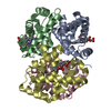
| ||||||||
|---|---|---|---|---|---|---|---|---|---|
| 1 |
| ||||||||
| Unit cell |
| ||||||||
| Noncrystallographic symmetry (NCS) | NCS oper: (Code: given Matrix: (0.213187, -0.332502, 0.918691), Vector: |
-Components
| #1: Protein | Mass: 15150.353 Da / Num. of mol.: 2 Source method: isolated from a genetically manipulated source Source: (gene. exp.) Homo sapiens (human) / References: UniProt: P69905#2: Protein | Mass: 15890.198 Da / Num. of mol.: 2 Source method: isolated from a genetically manipulated source Source: (gene. exp.) Homo sapiens (human) / References: UniProt: P68871#3: Chemical | ChemComp-HEM /   Mass: 616.487 Da / Num. of mol.: 4 / Source method: obtained synthetically / Formula: C34H32FeN4O4 Mass: 616.487 Da / Num. of mol.: 4 / Source method: obtained synthetically / Formula: C34H32FeN4O4#4: Chemical | ChemComp-CMO /   Mass: 28.010 Da / Num. of mol.: 4 / Source method: obtained synthetically / Formula: CO Mass: 28.010 Da / Num. of mol.: 4 / Source method: obtained synthetically / Formula: CO#5: Water | ChemComp-HOH / |  Mass: 18.015 Da / Num. of mol.: 302 / Source method: isolated from a natural source / Formula: H2O Mass: 18.015 Da / Num. of mol.: 302 / Source method: isolated from a natural source / Formula: H2OCompound details | PREVIOUS CRYSTALLOGRAPHIC STUDIES HAVE SHOWN THAT WILD-TYPE HUMAN HEMOGLOBIN CAN ADOPT TWO STABLE ...PREVIOUS CRYSTALLOG | |
|---|
-Experimental details
-Experiment
| Experiment | Method: X-RAY DIFFRACTION |
|---|
- Sample preparation
| Crystal | Density Matthews: 2.43 Å3/Da / Density % sol: 49.46 % | ||||||||||||||||||||||||
|---|---|---|---|---|---|---|---|---|---|---|---|---|---|---|---|---|---|---|---|---|---|---|---|---|---|
| Crystal grow | *PLUS Method: vapor diffusion, hanging drop | ||||||||||||||||||||||||
| Components of the solutions | *PLUS
|
-Data collection
| Radiation | Scattering type: x-ray |
|---|---|
| Radiation wavelength | Relative weight: 1 |
| Reflection | *PLUS Highest resolution: 1.7 Å / Num. measured all: 33563 |
- Processing
| Software | Name: PROLSQ / Classification: refinement | |||||||||||||||||||||||||||||||||||||||||||||||||||||||||||||||
|---|---|---|---|---|---|---|---|---|---|---|---|---|---|---|---|---|---|---|---|---|---|---|---|---|---|---|---|---|---|---|---|---|---|---|---|---|---|---|---|---|---|---|---|---|---|---|---|---|---|---|---|---|---|---|---|---|---|---|---|---|---|---|---|---|
| Refinement | Rfactor obs: 0.184 / Highest resolution: 1.7 Å | |||||||||||||||||||||||||||||||||||||||||||||||||||||||||||||||
| Refinement step | Cycle: LAST / Highest resolution: 1.7 Å
| |||||||||||||||||||||||||||||||||||||||||||||||||||||||||||||||
| Refine LS restraints |
| |||||||||||||||||||||||||||||||||||||||||||||||||||||||||||||||
| Software | *PLUS Name: PROLSQ / Classification: refinement | |||||||||||||||||||||||||||||||||||||||||||||||||||||||||||||||
| Refinement | *PLUS Highest resolution: 1.7 Å / Lowest resolution: 6 Å / Num. reflection obs: 71932 / σ(I): 2 / Rfactor obs: 0.184 | |||||||||||||||||||||||||||||||||||||||||||||||||||||||||||||||
| Solvent computation | *PLUS | |||||||||||||||||||||||||||||||||||||||||||||||||||||||||||||||
| Displacement parameters | *PLUS | |||||||||||||||||||||||||||||||||||||||||||||||||||||||||||||||
| Refine LS restraints | *PLUS
|
