 Movie
Movie Controller

[English] 日本語
 Yorodumi
- PDB-1hv4: CRYSTAL STRUCTURE ANALYSIS OF BAR-HEAD GOOSE HEMOGLOBIN (DEOXY FORM) -
+ Open data

- Basic information
| Entry | Database: PDB / ID: 1hv4 | ||||||
|---|---|---|---|---|---|---|---|
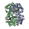
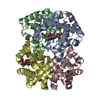
| Title | CRYSTAL STRUCTURE ANALYSIS OF BAR-HEAD GOOSE HEMOGLOBIN (DEOXY FORM) | ||||||
 Components Components |
| ||||||
 Keywords Keywords | OXYGEN STORAGE/TRANSPORT / ALLOSTERIC MECHANISM / OXYGEN TRANSPORT / HEME / RESPIRATORY PROTEIN / OXYGEN STORAGE-TRANSPORT COMPLEX | ||||||
| Function / homology |  Function and homology information Function and homology informationhaptoglobin binding / organic acid binding / haptoglobin-hemoglobin complex / hemoglobin complex / hydrogen peroxide catabolic process / oxygen carrier activity / peroxidase activity / oxygen binding / blood microparticle / iron ion binding ...haptoglobin binding / organic acid binding / haptoglobin-hemoglobin complex / hemoglobin complex / hydrogen peroxide catabolic process / oxygen carrier activity / peroxidase activity / oxygen binding / blood microparticle / iron ion binding / heme binding / metal ion binding Similarity search - Function | ||||||
| Biological species |  Anser indicus (Bar-headed goose) Anser indicus (Bar-headed goose) | ||||||
| Method |  X-RAY DIFFRACTION / MOLECULAR REPLACEMENT / Resolution: 2.8 Å X-RAY DIFFRACTION / MOLECULAR REPLACEMENT / Resolution: 2.8 Å | ||||||
 Authors Authors | Liang, Y. / Hua, Z. / Liang, X. / Xu, Q. / Lu, G. | ||||||
 Citation Citation | Journal: J.Mol.Biol. / Year: 2001 Title: The crystal structure of bar-headed goose hemoglobin in deoxy form: the allosteric mechanism of a hemoglobin species with high oxygen affinity. Authors: Liang, Y. / Hua, Z. / Liang, X. / Xu, Q. / Lu, G. | ||||||
| History |
|
- Structure visualization
| Structure viewer | Molecule: MolmilJmol/JSmol |
|---|
- Downloads & links
-Download
| PDBx/mmCIF format | 1hv4.cif.gz | 234.1 KB | Display | PDBx/mmCIF format |
|---|---|---|---|---|
| PDB format | pdb1hv4.ent.gz | 192.2 KB | Display | PDB format |
| PDBx/mmJSON format | 1hv4.json.gz | Tree view | PDBx/mmJSON format | |
| Others |  Other downloads Other downloads |
-Validation report
| Arichive directory | https://data.pdbj.org/pub/pdb/validation_reports/hv/1hv4ftp://data.pdbj.org/pub/pdb/validation_reports/hv/1hv4 | HTTPS FTP |
|---|
-Related structure data
| Related structure data |  1a4fS S: Starting model for refinement |
|---|---|
| Similar structure data |

-Links
 PDBj
PDBj


- Assembly
| Deposited unit | 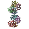
| ||||||||
|---|---|---|---|---|---|---|---|---|---|
| 1 | 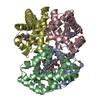
| ||||||||
| 2 | 
| ||||||||
| Unit cell |
|
-Components
| #1: Protein | Mass: 15361.679 Da / Num. of mol.: 4 / Source method: isolated from a natural source / Source: (natural) Anser indicus (Bar-headed goose) / Cell: ERYTHROCYTES / Cellular location: CYTOPLASM / Tissue: BLOOD / References: UniProt: P01990#2: Protein | Mass: 16313.866 Da / Num. of mol.: 4 / Source method: isolated from a natural source / Source: (natural) Anser indicus (Bar-headed goose) / Cell: ERYTHROCYTES / Cellular location: CYTOPLASM / Tissue: BLOOD / References: UniProt: P02118#3: Chemical | ChemComp-HEM /   Mass: 616.487 Da / Num. of mol.: 8 / Source method: obtained synthetically / Formula: C34H32FeN4O4 Mass: 616.487 Da / Num. of mol.: 8 / Source method: obtained synthetically / Formula: C34H32FeN4O4 |
|---|
-Experimental details
-Experiment
| Experiment | Method: X-RAY DIFFRACTION / Number of used crystals: 1 |
|---|
- Sample preparation
| Crystal | Density Matthews: 2.67 Å3/Da / Density % sol: 53.99 % | |||||||||||||||||||||||||
|---|---|---|---|---|---|---|---|---|---|---|---|---|---|---|---|---|---|---|---|---|---|---|---|---|---|---|
| Crystal grow | Temperature: 291 K / Method: vapor diffusion, hanging drop / pH: 7.2 Details: PEG 6000, potassium phosphate , pH 7.2, VAPOR DIFFUSION, HANGING DROP, temperature 291K | |||||||||||||||||||||||||
| Crystal grow | *PLUS Temperature: 18 ℃ | |||||||||||||||||||||||||
| Components of the solutions | *PLUS
|
-Data collection
| Diffraction | Mean temperature: 291 K |
|---|---|
| Diffraction source | Source: ROTATING ANODE / Type: RIGAKU RU300 / Wavelength: 1.5418 Å |
| Detector | Type: SIEMENS X200 / Detector: AREA DETECTOR / Date: May 10, 1995 |
| Radiation | Protocol: SINGLE WAVELENGTH / Monochromatic (M) / Laue (L): M / Scattering type: x-ray |
| Radiation wavelength | Wavelength: 1.5418 Å / Relative weight: 1 |
| Reflection | Resolution: 2.57→36.89 Å / Num. all: 45509 / Num. obs: 30654 / % possible obs: 67.4 % / Observed criterion σ(F): 1 / Observed criterion σ(I): 1 / Redundancy: 1.9 % / Biso Wilson estimate: 28.4 Å2 / Rmerge(I) obs: 0.061 / Net I/σ(I): 15.1 |
| Reflection shell | Resolution: 2.57→2.73 Å / Redundancy: 1.14 % / Rmerge(I) obs: 0.146 / Mean I/σ(I) obs: 4.4 / Num. unique all: 1779 / % possible all: 24 |
| Reflection | *PLUS Num. measured all: 57884 / Rmerge(I) obs: 0.0541 |
- Processing
| Software |
| |||||||||||||||||||||||||
|---|---|---|---|---|---|---|---|---|---|---|---|---|---|---|---|---|---|---|---|---|---|---|---|---|---|---|
| Refinement | Method to determine structure: MOLECULAR REPLACEMENT Starting model: PDB ENTRY 1A4F Resolution: 2.8→36.89 Å / Rfactor Rfree error: 0.005 / Data cutoff high absF: 63263.6 / Data cutoff low absF: 0 / Isotropic thermal model: RESTRAINED / Cross valid method: THROUGHOUT / σ(F): 0 / σ(I): 0 / Stereochemistry target values: Engh & Huber
| |||||||||||||||||||||||||
| Solvent computation | Solvent model: FLAT MODEL / Bsol: 30.63 Å2 / ksol: 0.299 e/Å3 | |||||||||||||||||||||||||
| Displacement parameters | Biso mean: 27.7 Å2
| |||||||||||||||||||||||||
| Refine analyze |
| |||||||||||||||||||||||||
| Refinement step | Cycle: LAST / Resolution: 2.8→36.89 Å
| |||||||||||||||||||||||||
| Refine LS restraints |
| |||||||||||||||||||||||||
| Refine LS restraints NCS | NCS model details: CONSTR | |||||||||||||||||||||||||
| LS refinement shell | Resolution: 2.8→2.98 Å / Rfactor Rfree error: 0.017 / Total num. of bins used: 6
| |||||||||||||||||||||||||
| Xplor file |
| |||||||||||||||||||||||||
| Software | *PLUS Name: CNS / Version: 1 / Classification: refinement | |||||||||||||||||||||||||
| Refinement | *PLUS σ(F): 0 / % reflection Rfree: 10.1 % | |||||||||||||||||||||||||
| Solvent computation | *PLUS | |||||||||||||||||||||||||
| Displacement parameters | *PLUS Biso mean: 27.7 Å2 | |||||||||||||||||||||||||
| Refine LS restraints | *PLUS
| |||||||||||||||||||||||||
| LS refinement shell | *PLUS Rfactor Rfree: 0.329 / % reflection Rfree: 10.6 % / Rfactor Rwork: 0.285 |
