 Movie
Movie Controller


+ Open data

- Basic information
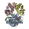
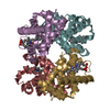
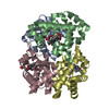
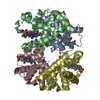
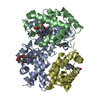
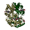
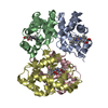
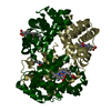
| Entry | Database: PDB / ID: 1jy7 | ||||||
|---|---|---|---|---|---|---|---|
| Title | THE STRUCTURE OF HUMAN METHEMOGLOBIN. THE VARIATION OF A THEME | ||||||
 Components Components |
| ||||||
 Keywords Keywords | OXYGEN STORAGE/TRANSPORT / HUMAN METHEMOGLOBIN / OXYGEN TRANSPORT / OXYGEN STORAGE-TRANSPORT COMPLEX | ||||||
| Function / homology |  Function and homology information Function and homology informationcellular oxidant detoxification / Heme assimilation / nitric oxide transport / hemoglobin alpha binding / hemoglobin binding / haptoglobin-hemoglobin complex / renal absorption / hemoglobin complex / oxygen transport / Scavenging of heme from plasma ...cellular oxidant detoxification / Heme assimilation / nitric oxide transport / hemoglobin alpha binding / hemoglobin binding / haptoglobin-hemoglobin complex / renal absorption / hemoglobin complex / oxygen transport / Scavenging of heme from plasma / erythrocyte development / endocytic vesicle lumen / blood vessel diameter maintenance / hydrogen peroxide catabolic process / oxygen carrier activity / carbon dioxide transport / response to hydrogen peroxide / Heme signaling / Erythrocytes take up oxygen and release carbon dioxide / Erythrocytes take up carbon dioxide and release oxygen / Cytoprotection by HMOX1 / oxygen binding / Late endosomal microautophagy / platelet aggregation / regulation of blood pressure / Chaperone Mediated Autophagy / positive regulation of nitric oxide biosynthetic process / tertiary granule lumen / Factors involved in megakaryocyte development and platelet production / blood microparticle / ficolin-1-rich granule lumen / iron ion binding / inflammatory response / heme binding / Neutrophil degranulation / : / extracellular exosome / extracellular region / membrane / metal ion binding / cytosol Similarity search - Function | ||||||
| Biological species |  Homo sapiens (human) Homo sapiens (human) | ||||||
| Method |  X-RAY DIFFRACTION / MOLECULAR REPLACEMENT / Resolution: 3.2 Å X-RAY DIFFRACTION / MOLECULAR REPLACEMENT / Resolution: 3.2 Å | ||||||
 Authors Authors | Biswal, B.K. / Vijayan, M. | ||||||
 Citation Citation | Journal: Acta Crystallogr.,Sect.D / Year: 2002 Title: Structures of human oxy- and deoxyhaemoglobin at different levels of humidity: variability in the T state. Authors: Biswal, B.K. / Vijayan, M. #1: Journal: Curr.Sci. / Year: 2001Title: Structure of human methaemoglobin: The variation of a theme Authors: Biswal, B.K. / Vijayan, M. #2: Journal: J.Mol.Biol. / Year: 1983Title: Structure of human oxyhaemoglobin at 2.1 A resolution Authors: Shaanan, B. #3: Journal: J.Mol.Biol. / Year: 1984Title: The Crystal Structure of Human Deoxyhemoglobin at 1.74 Angstroms Resolution Authors: Fermi, G. / Perutz, M.F. / Shaanan, B. / Fourme, R. #4: Journal: J.Biol.Chem. / Year: 1992Title: A third quaternary structure of human hemoglobin A at 1.7-A resolution Authors: Silva, M.M. / Rogers, P.H. / Arnone, A. | ||||||
| History |
|
- Structure visualization
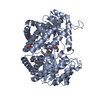
| Structure viewer | Molecule: MolmilJmol/JSmol |
|---|
- Downloads & links
-Download
| PDBx/mmCIF format | 1jy7.cif.gz | 307.4 KB | Display | PDBx/mmCIF format |
|---|---|---|---|---|
| PDB format | pdb1jy7.ent.gz | 260.2 KB | Display | PDB format |
| PDBx/mmJSON format | 1jy7.json.gz | Tree view | PDBx/mmJSON format | |
| Others |  Other downloads Other downloads |
-Validation report
| Arichive directory | https://data.pdbj.org/pub/pdb/validation_reports/jy/1jy7ftp://data.pdbj.org/pub/pdb/validation_reports/jy/1jy7 | HTTPS FTP |
|---|
-Related structure data
| Related structure data |  1lflC  1lfqC 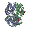 1lftC  1lfvC  1lfyC  1lfzC C: citing same article ( |
|---|---|
| Similar structure data |

-Links
 PDBj
PDBj


- Assembly
| Deposited unit | 
| ||||||||||
|---|---|---|---|---|---|---|---|---|---|---|---|
| 1 | 
| ||||||||||
| 2 | 
| ||||||||||
| 3 | 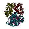
| ||||||||||
| Unit cell |
|
-Components
| #1: Protein | Mass: 15150.353 Da / Num. of mol.: 6 / Source method: isolated from a natural source / Source: (natural) Homo sapiens (human) / References: UniProt: P69905#2: Protein | Mass: 15890.198 Da / Num. of mol.: 6 / Source method: isolated from a natural source / Source: (natural) Homo sapiens (human) / References: UniProt: P68871#3: Chemical | ChemComp-HEM /   Mass: 616.487 Da / Num. of mol.: 12 / Source method: obtained synthetically / Formula: C34H32FeN4O4 Mass: 616.487 Da / Num. of mol.: 12 / Source method: obtained synthetically / Formula: C34H32FeN4O4 |
|---|
-Experimental details
-Experiment
| Experiment | Method: X-RAY DIFFRACTION / Number of used crystals: 1 |
|---|
- Sample preparation
| Crystal | Density Matthews: 2.53 Å3/Da / Density % sol: 51.4 % |
|---|---|
| Crystal grow | Temperature: 293 K / Method: liquid diffusion / pH: 6.7 Details: PEG 4000, Sodium potassium phosphate, pH 6.7, LIQUID DIFFUSION at 293K |
-Data collection
| Diffraction | Mean temperature: 293 K |
|---|---|
| Diffraction source | Source: ROTATING ANODE / Type: RIGAKU RU200 / Wavelength: 1.5418 Å |
| Detector | Type: RIGAKU / Detector: IMAGE PLATE |
| Radiation | Protocol: SINGLE WAVELENGTH / Monochromatic (M) / Laue (L): M / Scattering type: x-ray |
| Radiation wavelength | Wavelength: 1.5418 Å / Relative weight: 1 |
| Reflection | Resolution: 3.2→10 Å / Num. obs: 84268 / % possible obs: 92.7 % / Observed criterion σ(F): 0 / Observed criterion σ(I): 0 / Biso Wilson estimate: 38 Å2 / Rmerge(I) obs: 0.108 |
| Reflection shell | Resolution: 3.2→3.3 Å / Rmerge(I) obs: 0.379 |
- Processing
| Software |
| ||||||||||||||||||||
|---|---|---|---|---|---|---|---|---|---|---|---|---|---|---|---|---|---|---|---|---|---|
| Refinement | Method to determine structure: MOLECULAR REPLACEMENT Starting model: HUMAN OXYHEMOGLOBIN Resolution: 3.2→10 Å / Rfactor Rfree error: 0.008 / Data cutoff high absF: 158735.8 / Data cutoff low absF: 0 / Isotropic thermal model: RESTRAINED / Cross valid method: THROUGHOUT / σ(F): 2
| ||||||||||||||||||||
| Solvent computation | Solvent model: FLAT MODEL / Bsol: 51.9737 Å2 / ksol: 0.20873 e/Å3 | ||||||||||||||||||||
| Displacement parameters | Biso mean: 85 Å2
| ||||||||||||||||||||
| Refine analyze |
| ||||||||||||||||||||
| Refinement step | Cycle: LAST / Resolution: 3.2→10 Å
| ||||||||||||||||||||
| Refine LS restraints |
| ||||||||||||||||||||
| LS refinement shell | Resolution: 3.2→3.39 Å / Rfactor Rfree error: 0.043 / Total num. of bins used: 6
| ||||||||||||||||||||
| Xplor file |
|
