 Movie
Movie Controller

[English] 日本語
 Yorodumi
- PDB-3gys: Crystal structure determination of cat (Felis silvestris catus) h... -
+ Open data

- Basic information
| Entry | Database: PDB / ID: 3gys | ||||||
|---|---|---|---|---|---|---|---|
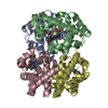
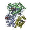
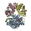
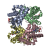
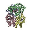
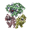
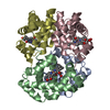
| Title | Crystal structure determination of cat (Felis silvestris catus) hemoglobin at 2.9 angstrom resolution | ||||||
 Components Components |
| ||||||
 Keywords Keywords | Oxygen storage / Oxygen transport / METHEMOGLOBIN / LOW-OXYGEN AFFINITY / ORTHORHOMBIC / chromatography / Heme / Iron / Transport / Polymorphism | ||||||
| Function / homology |  Function and homology information Function and homology informationhemoglobin alpha binding / haptoglobin-hemoglobin complex / hemoglobin complex / oxygen transport / erythrocyte development / oxygen carrier activity / oxygen binding / iron ion binding / heme binding / metal ion binding Similarity search - Function | ||||||
| Biological species |  | ||||||
| Method |  X-RAY DIFFRACTION / MOLECULAR REPLACEMENT / Resolution: 2.9 Å X-RAY DIFFRACTION / MOLECULAR REPLACEMENT / Resolution: 2.9 Å | ||||||
 Authors Authors | Balasubramanian, M. / Sathya Moorthy, P. / Neelagandan, K. / Ponnuswamy, M.N. | ||||||
 Citation Citation | Journal: To be Published Title: Crystal structure determination of cat (Felis silvestris catus) hemoglobin at 2.9 angstrom resolution Authors: Balasubramanian, M. / Sathya Moorthy, P. / Neelagandan, K. / Ponnuswamy, M.N. #1: Journal: Acta Crystallogr.,Sect.F / Year: 2007 Title: Crystallization of sheep (Ovis aries) and goat (Capra hircus) haemoglobins under unbuffered low-salt conditions Authors: Neelagandan, K. / Sathya Moorthy, P. / Balasubramanian, M. / Ponnuswamy, M.N. #2: Journal: Acta Crystallogr.,Sect.F / Year: 2009 Title: Purification, crystallization and preliminary crystallographic study of low oxygen-affinity haemoglobin from cat (Felis silvestris catus) in two different crystal forms Authors: Balasubramanian, M. / Sathya Moorthy, P. / Neelagandan, K. / Ponnuswamy, M.N. #3: Journal: Protein Pept.Lett. / Year: 2009 Title: Preliminary Crystallographic Study of Hemoglobin from Buffalo (Bubalus bubalis): A Low Oxygen Affinity Species Authors: Balasubramanian, M. / Sathya Moorthy, P. / Neelagandan, K. / Ponnuswamy, M.N. #4: Journal: Protein Pept.Lett. / Year: 2009Title: Purification, crystallization and preliminary X-ray diffraction studies on goat (Capra hircus) hemoglobin - a low oxygen affinity species Authors: Sathya Moorthy, P. / Neelagandan, K. / Balasubramanian, M. / Ponnuswamy, M.N. | ||||||
| History |
|
- Structure visualization
| Structure viewer | Molecule: MolmilJmol/JSmol |
|---|
- Downloads & links
-Download
| PDBx/mmCIF format | 3gys.cif.gz | 228.6 KB | Display | PDBx/mmCIF format |
|---|---|---|---|---|
| PDB format | pdb3gys.ent.gz | 187.7 KB | Display | PDB format |
| PDBx/mmJSON format | 3gys.json.gz | Tree view | PDBx/mmJSON format | |
| Others |  Other downloads Other downloads |
-Validation report
| Arichive directory | https://data.pdbj.org/pub/pdb/validation_reports/gy/3gysftp://data.pdbj.org/pub/pdb/validation_reports/gy/3gys | HTTPS FTP |
|---|
-Related structure data
| Related structure data |  3d4xS S: Starting model for refinement |
|---|---|
| Similar structure data |


-Links
 PDBj
PDBj


- Assembly
| Deposited unit | 
| ||||||||
|---|---|---|---|---|---|---|---|---|---|
| 1 | 
| ||||||||
| 2 | 
| ||||||||
| 3 | 
| ||||||||
| 4 | 
| ||||||||
| 5 | 
| ||||||||
| 6 | 
| ||||||||
| Unit cell |
|
-Components
| #1: Protein | Mass: 15328.458 Da / Num. of mol.: 4 / Source method: isolated from a natural source / Source: (natural) #2: Protein | Mass: 15892.134 Da / Num. of mol.: 4 / Source method: isolated from a natural source / Source: (natural) #3: Chemical | ChemComp-HEM /   Mass: 616.487 Da / Num. of mol.: 8 / Source method: obtained synthetically / Formula: C34H32FeN4O4 Mass: 616.487 Da / Num. of mol.: 8 / Source method: obtained synthetically / Formula: C34H32FeN4O4#4: Water | ChemComp-HOH / |  Mass: 18.015 Da / Num. of mol.: 9 / Source method: isolated from a natural source / Formula: H2O Mass: 18.015 Da / Num. of mol.: 9 / Source method: isolated from a natural source / Formula: H2O |
|---|
-Experimental details
-Experiment
| Experiment | Method: X-RAY DIFFRACTION / Number of used crystals: 1 |
|---|
- Sample preparation
| Crystal | Density Matthews: 2.43 Å3/Da / Density % sol: 49.4 % |
|---|---|
| Crystal grow | Temperature: 293 K / Method: vapor diffusion, hanging drop / pH: 6.7 Details: 40% PEG 3350, 50mM phosphate buffer at pH 6.7, 1M NaCl, VAPOR DIFFUSION, HANGING DROP, temperature 293K |
-Data collection
| Diffraction | Mean temperature: 293 K |
|---|---|
| Diffraction source | Source: ROTATING ANODE / Type: RIGAKU RU300 / Wavelength: 1.5417 Å |
| Detector | Type: MAR scanner 345 mm plate / Detector: IMAGE PLATE / Date: Oct 12, 2007 |
| Radiation | Monochromator: MIRROS / Protocol: SINGLE WAVELENGTH / Monochromatic (M) / Laue (L): M / Scattering type: x-ray |
| Radiation wavelength | Wavelength: 1.5417 Å / Relative weight: 1 |
| Reflection | Resolution: 2.9→30 Å / Num. all: 26800 / Num. obs: 23886 / % possible obs: 96 % / Observed criterion σ(F): 2 / Observed criterion σ(I): 2 / Redundancy: 3.65 % / Rsym value: 0.098 / Net I/σ(I): 4.1 |
| Reflection shell | Resolution: 2.9→3 Å / Redundancy: 3.14 % / Mean I/σ(I) obs: 1.4 / Num. unique all: 2304 / Rsym value: 0.2648 / % possible all: 98.5 |
- Processing
| Software |
| |||||||||||||||||||||||||||||||||||||||||||||||||||
|---|---|---|---|---|---|---|---|---|---|---|---|---|---|---|---|---|---|---|---|---|---|---|---|---|---|---|---|---|---|---|---|---|---|---|---|---|---|---|---|---|---|---|---|---|---|---|---|---|---|---|---|---|
| Refinement | Method to determine structure: MOLECULAR REPLACEMENT Starting model: PDB ENTRY 3D4X Resolution: 2.9→24.13 Å / Cor.coef. Fo:Fc: 0.947 / Cor.coef. Fo:Fc free: 0.889 / SU B: 20.016 / SU ML: 0.378 / Cross valid method: THROUGHOUT / σ(F): 2 / σ(I): 2 / ESU R Free: 0.508 / Stereochemistry target values: MAXIMUM LIKELIHOOD
| |||||||||||||||||||||||||||||||||||||||||||||||||||
| Solvent computation | Ion probe radii: 0.8 Å / Shrinkage radii: 0.8 Å / VDW probe radii: 1.2 Å / Solvent model: MASK | |||||||||||||||||||||||||||||||||||||||||||||||||||
| Displacement parameters | Biso mean: 48.397 Å2
| |||||||||||||||||||||||||||||||||||||||||||||||||||
| Refinement step | Cycle: LAST / Resolution: 2.9→24.13 Å
| |||||||||||||||||||||||||||||||||||||||||||||||||||
| Refine LS restraints |
| |||||||||||||||||||||||||||||||||||||||||||||||||||
| LS refinement shell | Resolution: 2.9→2.975 Å / Total num. of bins used: 20
|
