 Movie
Movie Controller


+ Open data

- Basic information
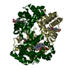
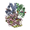
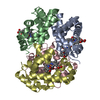
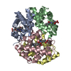
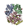
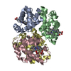
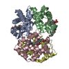
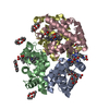
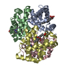
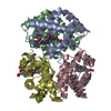
| Entry | Database: PDB / ID: 1buw | ||||||
|---|---|---|---|---|---|---|---|
| Title | CRYSTAL STRUCTURE OF S-NITROSO-NITROSYL HUMAN HEMOGLOBIN A | ||||||
 Components Components | (PROTEIN (HEMOGLOBIN)) x 2 | ||||||
 Keywords Keywords | OXYGEN STORAGE/TRANSPORT / OXYGEN TRANSPORT AND VASODILATION / OXYGEN STORAGE-TRANSPORT COMPLEX | ||||||
| Function / homology |  Function and homology information Function and homology information: / haptoglobin binding / cellular oxidant detoxification / Heme assimilation / nitric oxide transport / hemoglobin alpha binding / hemoglobin binding / haptoglobin-hemoglobin complex / renal absorption / hemoglobin complex ...: / haptoglobin binding / cellular oxidant detoxification / Heme assimilation / nitric oxide transport / hemoglobin alpha binding / hemoglobin binding / haptoglobin-hemoglobin complex / renal absorption / hemoglobin complex / oxygen transport / Scavenging of heme from plasma / erythrocyte development / endocytic vesicle lumen / blood vessel diameter maintenance / hydrogen peroxide catabolic process / oxygen carrier activity / peroxidase activity / carbon dioxide transport / response to hydrogen peroxide / Heme signaling / Erythrocytes take up oxygen and release carbon dioxide / Erythrocytes take up carbon dioxide and release oxygen / Cytoprotection by HMOX1 / oxygen binding / Late endosomal microautophagy / platelet aggregation / regulation of blood pressure / Chaperone Mediated Autophagy / positive regulation of nitric oxide biosynthetic process / tertiary granule lumen / Factors involved in megakaryocyte development and platelet production / blood microparticle / ficolin-1-rich granule lumen / iron ion binding / inflammatory response / heme binding / Neutrophil degranulation / : / extracellular exosome / extracellular region / membrane / metal ion binding / cytosol Similarity search - Function | ||||||
| Biological species |  Homo sapiens (human) Homo sapiens (human) | ||||||
| Method |  X-RAY DIFFRACTION / OTHER / Resolution: 1.9 Å X-RAY DIFFRACTION / OTHER / Resolution: 1.9 Å | ||||||
 Authors Authors | Chan, N.-L. / Rogers, P.H. / Arnone, A. | ||||||
 Citation Citation | Journal: Biochemistry / Year: 1998 Title: Crystal structure of the S-nitroso form of liganded human hemoglobin. Authors: Chan, N.L. / Rogers, P.H. / Arnone, A. | ||||||
| History |
|
- Structure visualization
| Structure viewer | Molecule: MolmilJmol/JSmol |
|---|
- Downloads & links
-Download
| PDBx/mmCIF format | 1buw.cif.gz | 126.2 KB | Display | PDBx/mmCIF format |
|---|---|---|---|---|
| PDB format | pdb1buw.ent.gz | 100.5 KB | Display | PDB format |
| PDBx/mmJSON format | 1buw.json.gz | Tree view | PDBx/mmJSON format | |
| Others |  Other downloads Other downloads |
-Validation report
| Arichive directory | https://data.pdbj.org/pub/pdb/validation_reports/bu/1buwftp://data.pdbj.org/pub/pdb/validation_reports/bu/1buw | HTTPS FTP |
|---|
-Related structure data
| Related structure data | 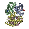 1bbbS S: Starting model for refinement |
|---|---|
| Similar structure data |

-Links
 PDBj
PDBj


- Assembly
| Deposited unit | 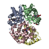
| ||||||||
|---|---|---|---|---|---|---|---|---|---|
| 1 |
| ||||||||
| Unit cell |
| ||||||||
| Noncrystallographic symmetry (NCS) | NCS oper: (Code: given Matrix: (0.202601, -0.326635, 0.923181), Vector: |
-Components
| #1: Protein | Mass: 15150.353 Da / Num. of mol.: 2 / Source method: isolated from a natural source / Details: OUDATED BLOOD DRAWN FROM HEALTHY NON-SMOKER. / Source: (natural) Homo sapiens (human) / Cell: RED BLOOD CELL / References: UniProt: P69905#2: Protein | Mass: 15919.196 Da / Num. of mol.: 2 / Source method: isolated from a natural source / Details: OUDATED BLOOD DRAWN FROM HEALTHY NON-SMOKER. / Source: (natural) Homo sapiens (human) / Cell: RED BLOOD CELL / References: UniProt: P02023, UniProt: P68871*PLUS#3: Chemical | ChemComp-HEM /   Mass: 616.487 Da / Num. of mol.: 4 / Source method: obtained synthetically / Formula: C34H32FeN4O4 Mass: 616.487 Da / Num. of mol.: 4 / Source method: obtained synthetically / Formula: C34H32FeN4O4#4: Chemical | ChemComp-NO /   Mass: 30.006 Da / Num. of mol.: 4 / Source method: obtained synthetically / Formula: NO Mass: 30.006 Da / Num. of mol.: 4 / Source method: obtained synthetically / Formula: NO#5: Water | ChemComp-HOH / |  Mass: 18.015 Da / Num. of mol.: 196 / Source method: isolated from a natural source / Formula: H2O Mass: 18.015 Da / Num. of mol.: 196 / Source method: isolated from a natural source / Formula: H2OCompound details | THIS STRUCTURE PROVIDES THE FIRST DIRECT VISUALIZAT | Has protein modification | Y | |
|---|
-Experimental details
-Experiment
| Experiment | Method: X-RAY DIFFRACTION / Number of used crystals: 3 |
|---|
- Sample preparation
| Crystal | Density Matthews: 2.4 Å3/Da / Density % sol: 49.06 % | ||||||||||||||||||||||||
|---|---|---|---|---|---|---|---|---|---|---|---|---|---|---|---|---|---|---|---|---|---|---|---|---|---|
| Crystal grow | pH: 5.8 Details: 1% PROTEIN 100 MM SODIUM CACODYLATE, PH 5.8 16% PEG 6000 | ||||||||||||||||||||||||
| Crystal grow | *PLUS Method: vapor diffusion, hanging drop / Details: Silva, M.M., (1992) J. Biol. Chem., 267, 17248. | ||||||||||||||||||||||||
| Components of the solutions | *PLUS
|
-Data collection
| Diffraction | Mean temperature: 293 K |
|---|---|
| Diffraction source | Source: ROTATING ANODE / Type: RIGAKU RU200 / Wavelength: 1.5418 |
| Detector | Type: SDMS / Detector: AREA DETECTOR / Date: Apr 15, 1997 |
| Radiation | Protocol: SINGLE WAVELENGTH / Monochromatic (M) / Laue (L): M / Scattering type: x-ray |
| Radiation wavelength | Wavelength: 1.5418 Å / Relative weight: 1 |
| Reflection | Resolution: 1.77→20 Å / Num. obs: 55671 / % possible obs: 93 % / Observed criterion σ(I): 2 / Redundancy: 4.9 % / Rsym value: 0.07 / Net I/σ(I): 17 |
| Reflection shell | Resolution: 1.77→1.9 Å / Redundancy: 2.5 % / Mean I/σ(I) obs: 2.5 / Rsym value: 0.25 / % possible all: 77 |
| Reflection | *PLUS % possible obs: 93 % / Num. measured all: 273695 / Rmerge(I) obs: 0.07 |
- Processing
| Software |
| ||||||||||||||||||||||||||||||||||||||||||||||||||||||||||||
|---|---|---|---|---|---|---|---|---|---|---|---|---|---|---|---|---|---|---|---|---|---|---|---|---|---|---|---|---|---|---|---|---|---|---|---|---|---|---|---|---|---|---|---|---|---|---|---|---|---|---|---|---|---|---|---|---|---|---|---|---|---|
| Refinement | Method to determine structure: OTHER Starting model: PDB ENTRY 1BBB Resolution: 1.9→8 Å / Cross valid method: THROUGHOUT / σ(F): 2
| ||||||||||||||||||||||||||||||||||||||||||||||||||||||||||||
| Refinement step | Cycle: LAST / Resolution: 1.9→8 Å
| ||||||||||||||||||||||||||||||||||||||||||||||||||||||||||||
| Refine LS restraints |
| ||||||||||||||||||||||||||||||||||||||||||||||||||||||||||||
| Xplor file |
| ||||||||||||||||||||||||||||||||||||||||||||||||||||||||||||
| Software | *PLUS Version: 3.1 / Classification: refinement | ||||||||||||||||||||||||||||||||||||||||||||||||||||||||||||
| Refinement | *PLUS Highest resolution: 1.9 Å / Lowest resolution: 8 Å / σ(F): 2 / % reflection Rfree: 10 % | ||||||||||||||||||||||||||||||||||||||||||||||||||||||||||||
| Solvent computation | *PLUS | ||||||||||||||||||||||||||||||||||||||||||||||||||||||||||||
| Displacement parameters | *PLUS | ||||||||||||||||||||||||||||||||||||||||||||||||||||||||||||
| Refine LS restraints | *PLUS
|
