 Movie
Movie Controller

[English] 日本語
 Yorodumi
- PDB-1rqa: Crystallographic Analysis of the Interaction of Nitric Oxide with... -
+ Open data

- Basic information
| Entry | Database: PDB / ID: 1rqa | ||||||
|---|---|---|---|---|---|---|---|
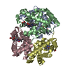
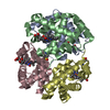
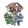
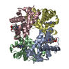
| Title | Crystallographic Analysis of the Interaction of Nitric Oxide with Quaternary-T Human Hemoglobin. Beta W73E hemoglobin exposed to NO under anaerobic conditions | ||||||
 Components Components |
| ||||||
 Keywords Keywords | OXYGEN STORAGE/TRANSPORT / globin / OXYGEN STORAGE-TRANSPORT COMPLEX | ||||||
| Function / homology |  Function and homology information Function and homology informationcellular oxidant detoxification / Heme assimilation / nitric oxide transport / hemoglobin alpha binding / hemoglobin binding / haptoglobin-hemoglobin complex / renal absorption / hemoglobin complex / oxygen transport / Scavenging of heme from plasma ...cellular oxidant detoxification / Heme assimilation / nitric oxide transport / hemoglobin alpha binding / hemoglobin binding / haptoglobin-hemoglobin complex / renal absorption / hemoglobin complex / oxygen transport / Scavenging of heme from plasma / erythrocyte development / endocytic vesicle lumen / blood vessel diameter maintenance / hydrogen peroxide catabolic process / oxygen carrier activity / carbon dioxide transport / response to hydrogen peroxide / Heme signaling / Erythrocytes take up oxygen and release carbon dioxide / Erythrocytes take up carbon dioxide and release oxygen / Cytoprotection by HMOX1 / oxygen binding / Late endosomal microautophagy / platelet aggregation / regulation of blood pressure / Chaperone Mediated Autophagy / positive regulation of nitric oxide biosynthetic process / tertiary granule lumen / Factors involved in megakaryocyte development and platelet production / blood microparticle / ficolin-1-rich granule lumen / iron ion binding / inflammatory response / heme binding / Neutrophil degranulation / : / extracellular exosome / extracellular region / membrane / metal ion binding / cytosol Similarity search - Function | ||||||
| Biological species |  Homo sapiens (human) Homo sapiens (human) | ||||||
| Method |  X-RAY DIFFRACTION / MOLECULAR REPLACEMENT / Resolution: 2.11 Å X-RAY DIFFRACTION / MOLECULAR REPLACEMENT / Resolution: 2.11 Å | ||||||
 Authors Authors | Chan, N.-L. / Kavanaugh, J.S. / Rogers, P.H. / Arnone, A. | ||||||
 Citation Citation | Journal: Biochemistry / Year: 2004 Title: Crystallographic analysis of the interaction of nitric oxide with quaternary-T human hemoglobin. Authors: Chan, N.-L. / Kavanaugh, J.S. / Rogers, P.H. / Arnone, A. #1: Journal: To be publishedTitle: X-ray Diffraction Studies of Fully Liganded 37beta Mutant Hemoglobins Reveal a T-Like Intermediate Structure with High Ligand Affinity Authors: Kavanaugh, J.S. / Rogers, P.H. / Arnone, A. | ||||||
| History |
|
- Structure visualization
| Structure viewer | Molecule: MolmilJmol/JSmol |
|---|
- Downloads & links
-Download
| PDBx/mmCIF format | 1rqa.cif.gz | 126.4 KB | Display | PDBx/mmCIF format |
|---|---|---|---|---|
| PDB format | pdb1rqa.ent.gz | 100.2 KB | Display | PDB format |
| PDBx/mmJSON format | 1rqa.json.gz | Tree view | PDBx/mmJSON format | |
| Others |  Other downloads Other downloads |
-Validation report
| Arichive directory | https://data.pdbj.org/pub/pdb/validation_reports/rq/1rqaftp://data.pdbj.org/pub/pdb/validation_reports/rq/1rqa | HTTPS FTP |
|---|
-Related structure data


-Links
 PDBj
PDBj


- Assembly
| Deposited unit | 
| ||||||||
|---|---|---|---|---|---|---|---|---|---|
| 1 |
| ||||||||
| Unit cell |
|
-Components
| #1: Protein | Mass: 15150.353 Da / Num. of mol.: 2 / Source method: isolated from a natural source Details: alpha globin subunits are purified from red blood cells Source: (natural) Homo sapiens (human) / Tissue: blood / References: UniProt: P69905#2: Protein | Mass: 15865.167 Da / Num. of mol.: 2 / Mutation: W37E, V1M Source method: isolated from a genetically manipulated source Source: (gene. exp.) Homo sapiens (human)Description: Tetramers are reconsituted by combining the mutant beta globin, heme and alpha subunits purified from red blood cells Gene: HBB / Production host:  #3: Chemical | ChemComp-HEM /   Mass: 616.487 Da / Num. of mol.: 4 / Source method: obtained synthetically / Formula: C34H32FeN4O4 Mass: 616.487 Da / Num. of mol.: 4 / Source method: obtained synthetically / Formula: C34H32FeN4O4#4: Chemical | ChemComp-NO /   Mass: 30.006 Da / Num. of mol.: 4 / Source method: obtained synthetically / Formula: NO Mass: 30.006 Da / Num. of mol.: 4 / Source method: obtained synthetically / Formula: NO#5: Water | ChemComp-HOH / |  Mass: 18.015 Da / Num. of mol.: 138 / Source method: isolated from a natural source / Formula: H2O Mass: 18.015 Da / Num. of mol.: 138 / Source method: isolated from a natural source / Formula: H2O |
|---|
-Experimental details
-Experiment
| Experiment | Method: X-RAY DIFFRACTION / Number of used crystals: 1 |
|---|
- Sample preparation
| Crystal | Density Matthews: 2.51 Å3/Da / Density % sol: 50.98 % |
|---|---|
| Crystal grow | Temperature: 298 K / pH: 7 Details: 10 mg/mL human hemoglobin, 10 mM potassium phosphate, 100 mM potassium chloride, 10-12% PEG 6000, 3mM sodium dithionite (under anaerobic conditions), pH 7.0, BATCH METHOD, temperature 298K |
-Data collection
| Diffraction | Mean temperature: 298 K |
|---|---|
| Diffraction source | Source: ROTATING ANODE / Type: RIGAKU RU200 / Wavelength: 1.5418 Å |
| Detector | Type: SDMS / Detector: AREA DETECTOR / Date: Sep 23, 1998 |
| Radiation | Monochromator: GRAPHITE / Protocol: SINGLE WAVELENGTH / Monochromatic (M) / Laue (L): M / Scattering type: x-ray |
| Radiation wavelength | Wavelength: 1.5418 Å / Relative weight: 1 |
| Reflection | Resolution: 2.11→55.048 Å / Num. obs: 42912 / % possible obs: 98 % / Redundancy: 6.6 % / Rmerge(I) obs: 0.085 / Net I/σ(I): 9.6 |
| Reflection shell | Resolution: 2.11→2.27 Å / Redundancy: 2.6 % / Rmerge(I) obs: 0.231 / Mean I/σ(I) obs: 2 / % possible all: 90.5 |
- Processing
| Software |
| ||||||||||||||||||||||||||||||||||||||||||||||||||||||||||||||||||||||||||||||||||||||||||||||||||||||||||||||||||||||||||||||||||||||||||||||||||||||||||||||||
|---|---|---|---|---|---|---|---|---|---|---|---|---|---|---|---|---|---|---|---|---|---|---|---|---|---|---|---|---|---|---|---|---|---|---|---|---|---|---|---|---|---|---|---|---|---|---|---|---|---|---|---|---|---|---|---|---|---|---|---|---|---|---|---|---|---|---|---|---|---|---|---|---|---|---|---|---|---|---|---|---|---|---|---|---|---|---|---|---|---|---|---|---|---|---|---|---|---|---|---|---|---|---|---|---|---|---|---|---|---|---|---|---|---|---|---|---|---|---|---|---|---|---|---|---|---|---|---|---|---|---|---|---|---|---|---|---|---|---|---|---|---|---|---|---|---|---|---|---|---|---|---|---|---|---|---|---|---|---|---|---|---|
| Refinement | Method to determine structure: MOLECULAR REPLACEMENT Starting model: betaW37E deoxyhemoglobin to be deposited in PDB Resolution: 2.11→10 Å / Cor.coef. Fo:Fc: 0.949 / Cor.coef. Fo:Fc free: 0.927 / SU B: 7.417 / SU ML: 0.195 / Cross valid method: THROUGHOUT / σ(F): 0 / ESU R: 0.243 / ESU R Free: 0.193 / Stereochemistry target values: MAXIMUM LIKELIHOOD / Details: HYDROGENS HAVE BEEN ADDED IN THE RIDING POSITIONS
| ||||||||||||||||||||||||||||||||||||||||||||||||||||||||||||||||||||||||||||||||||||||||||||||||||||||||||||||||||||||||||||||||||||||||||||||||||||||||||||||||
| Solvent computation | Ion probe radii: 0.8 Å / Shrinkage radii: 0.8 Å / VDW probe radii: 1.4 Å / Solvent model: BABINET MODEL WITH MASK | ||||||||||||||||||||||||||||||||||||||||||||||||||||||||||||||||||||||||||||||||||||||||||||||||||||||||||||||||||||||||||||||||||||||||||||||||||||||||||||||||
| Displacement parameters | Biso mean: 24.375 Å2
| ||||||||||||||||||||||||||||||||||||||||||||||||||||||||||||||||||||||||||||||||||||||||||||||||||||||||||||||||||||||||||||||||||||||||||||||||||||||||||||||||
| Refinement step | Cycle: LAST / Resolution: 2.11→10 Å
| ||||||||||||||||||||||||||||||||||||||||||||||||||||||||||||||||||||||||||||||||||||||||||||||||||||||||||||||||||||||||||||||||||||||||||||||||||||||||||||||||
| Refine LS restraints |
|
