 Movie
Movie Controller

[English] 日本語
 Yorodumi
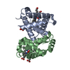
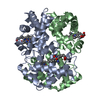
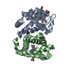
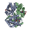
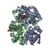
- PDB-2hbf: HIGH RESOLUTION X-RAY STRUCTURES OF MYOGLOBIN-AND HEMOGLOBIN-ALKY... -
+ Open data

- Basic information
| Entry | Database: PDB / ID: 2hbf | ||||||
|---|---|---|---|---|---|---|---|
| Title | HIGH RESOLUTION X-RAY STRUCTURES OF MYOGLOBIN-AND HEMOGLOBIN-ALKYL ISOCYANIDE COMPLEXES | ||||||
 Components Components |
| ||||||
 Keywords Keywords | OXYGEN TRANSPORT | ||||||
| Function / homology |  Function and homology information Function and homology informationcellular oxidant detoxification / Heme assimilation / nitric oxide transport / hemoglobin alpha binding / hemoglobin binding / haptoglobin-hemoglobin complex / renal absorption / hemoglobin complex / oxygen transport / Scavenging of heme from plasma ...cellular oxidant detoxification / Heme assimilation / nitric oxide transport / hemoglobin alpha binding / hemoglobin binding / haptoglobin-hemoglobin complex / renal absorption / hemoglobin complex / oxygen transport / Scavenging of heme from plasma / erythrocyte development / endocytic vesicle lumen / blood vessel diameter maintenance / hydrogen peroxide catabolic process / oxygen carrier activity / carbon dioxide transport / response to hydrogen peroxide / Heme signaling / Erythrocytes take up oxygen and release carbon dioxide / Erythrocytes take up carbon dioxide and release oxygen / Cytoprotection by HMOX1 / oxygen binding / Late endosomal microautophagy / platelet aggregation / regulation of blood pressure / Chaperone Mediated Autophagy / positive regulation of nitric oxide biosynthetic process / tertiary granule lumen / Factors involved in megakaryocyte development and platelet production / blood microparticle / ficolin-1-rich granule lumen / iron ion binding / inflammatory response / heme binding / Neutrophil degranulation / : / extracellular exosome / extracellular region / membrane / metal ion binding / cytosol Similarity search - Function | ||||||
| Biological species |  Homo sapiens (human) Homo sapiens (human) | ||||||
| Method |  X-RAY DIFFRACTION / Resolution: 2.2 Å X-RAY DIFFRACTION / Resolution: 2.2 Å | ||||||
 Authors Authors | Johnson, K.A. / Olson, J.S. / Phillips Jr., G.N. | ||||||
 Citation Citation | Journal: Thesis / Year: 1993 Title: High Resolution X-Ray Structures of Myoglobin-and Hemoglobin-Alkyl Isocyanide Complexes Authors: Johnson, K.A. #1: Journal: J.Mol.Biol. / Year: 1989Title: Structure of Myoglobin-Ethyl Isocyanide: Histidine as a Swinging Door for Ligand Entry Authors: Johnson, K.A. / Olson, J.S. / Phillips Jr., G.N. | ||||||
| History |
|
- Structure visualization
| Structure viewer | Molecule: MolmilJmol/JSmol |
|---|
- Downloads & links
-Download
| PDBx/mmCIF format | 2hbf.cif.gz | 70 KB | Display | PDBx/mmCIF format |
|---|---|---|---|---|
| PDB format | pdb2hbf.ent.gz | 53.4 KB | Display | PDB format |
| PDBx/mmJSON format | 2hbf.json.gz | Tree view | PDBx/mmJSON format | |
| Others |  Other downloads Other downloads |
-Validation report
| Arichive directory | https://data.pdbj.org/pub/pdb/validation_reports/hb/2hbfftp://data.pdbj.org/pub/pdb/validation_reports/hb/2hbf | HTTPS FTP |
|---|
-Related structure data
| Similar structure data |
|---|


-Links
 PDBj
PDBj


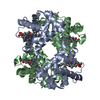
- Assembly
| Deposited unit | 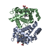
| ||||||||
|---|---|---|---|---|---|---|---|---|---|
| 1 | 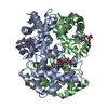
| ||||||||
| Unit cell |
|
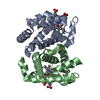
-Components
| #1: Protein | Mass: 15150.353 Da / Num. of mol.: 1 Source method: isolated from a genetically manipulated source Source: (gene. exp.) Homo sapiens (human) / Organ: SEED / References: UniProt: P69905 | ||||
|---|---|---|---|---|---|
| #2: Protein | Mass: 15890.198 Da / Num. of mol.: 1 Source method: isolated from a genetically manipulated source Source: (gene. exp.) Homo sapiens (human) / Organ: SEED / References: UniProt: P68871 | ||||
| #3: Chemical |   Mass: 616.487 Da / Num. of mol.: 2 / Source method: obtained synthetically / Formula: C34H32FeN4O4 Mass: 616.487 Da / Num. of mol.: 2 / Source method: obtained synthetically / Formula: C34H32FeN4O4#4: Chemical |   Mass: 69.105 Da / Num. of mol.: 2 / Source method: obtained synthetically / Formula: C4H7N Mass: 69.105 Da / Num. of mol.: 2 / Source method: obtained synthetically / Formula: C4H7N#5: Water | ChemComp-HOH / |  Mass: 18.015 Da / Num. of mol.: 55 / Source method: isolated from a natural source / Formula: H2O Mass: 18.015 Da / Num. of mol.: 55 / Source method: isolated from a natural source / Formula: H2O |
-Experimental details
-Experiment
| Experiment | Method: X-RAY DIFFRACTION |
|---|
- Sample preparation
| Crystal | Density Matthews: 2.28 Å3/Da / Density % sol: 46.03 % |
|---|
- Processing
| Software |
| ||||||||||||||||||||||||||||||||||||||||||||||||||||||||||||||||||||||||||||||||
|---|---|---|---|---|---|---|---|---|---|---|---|---|---|---|---|---|---|---|---|---|---|---|---|---|---|---|---|---|---|---|---|---|---|---|---|---|---|---|---|---|---|---|---|---|---|---|---|---|---|---|---|---|---|---|---|---|---|---|---|---|---|---|---|---|---|---|---|---|---|---|---|---|---|---|---|---|---|---|---|---|---|
| Refinement | Resolution: 2.2→5 Å / Rfactor Rwork: 0.179 / Rfactor obs: 0.179 / σ(F): 0 Details: REFINEMENT. THE STARTING MODEL WAS THE ETHYL ISOCYANIDE - HEMOGLOBIN COMPLEX WITH THE LIGAND REMOVED AND THE N-PROPYL ISOCYANIDE LIGAND MODELED INTO INITIAL DIFFERENCE MAP (FO-FC) ELECTRON ...Details: REFINEMENT. THE STARTING MODEL WAS THE ETHYL ISOCYANIDE - HEMOGLOBIN COMPLEX WITH THE LIGAND REMOVED AND THE N-PROPYL ISOCYANIDE LIGAND MODELED INTO INITIAL DIFFERENCE MAP (FO-FC) ELECTRON DENSITY. THE PROGRAM X-PLOR WAS USED TO ACHIEVE A FINAL CONVENTIONAL R-FACTOR OF 17.9%. THE REFINEMENT PROCESS INCLUDED SIMULATED ANNEALING FOLLOWED BY POSITION AND TEMPERATURE FACTOR REFINEMENT. AN ITERATIVE PROCESS OF MANUAL REFITTING OF SIDE CHAINS AND PLACEMENT OF WATERS WAS PERFORMED UNTIL THE R-FACTOR CONVERGED. THE WEIGHTING OF PSEUDO-ENERGY X-RAY TERM WAS ADJUSTED TO GIVE A BOND RMS OF 0.020 ANGSTROMS IN THE LAST FEW STEPS OF POSITIONAL REFINEMENT. WATERS (N=109) WERE RETAINED FROM THE STARTING STRUCTURE OF OXYHEMOGLOBIN (1HHO). ADDITIONAL WATERS WERE ADDED IF THEY LAY IN 3.5 - 4.O SIGMA PEAKS IN FO-FC ELECTRON DENSITY MAPS AND 1 SIGMA PEAKS IN 2FO-FC MAPS. CONCURRENTLY, A WATER WOULD BE DELETED IF ITS OCCUPANCY (Q) AND TEMPERATURE FACTOR (B) COMBINED TO MAKE THE VALUE (Q*EXP(- B/36)*100%) FALL BELOW 10%. A PEAK NUMBER OF 114 WATERS WAS REACHED. THIS WAS REDUCED TO 55 WATERS OVER THE LAST FEW REFINEMENT CYCLES BY DELETING WATERS WHICH FELL BELOW A 20% THRESHOLD. | ||||||||||||||||||||||||||||||||||||||||||||||||||||||||||||||||||||||||||||||||
| Refinement step | Cycle: LAST / Resolution: 2.2→5 Å
| ||||||||||||||||||||||||||||||||||||||||||||||||||||||||||||||||||||||||||||||||
| Refine LS restraints |
|
