 Movie
Movie Controller

[English] 日本語
 Yorodumi
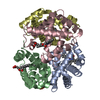
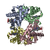
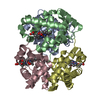
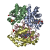
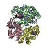
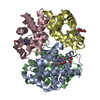
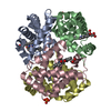
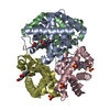
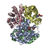
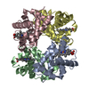
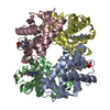
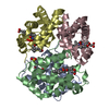
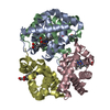
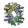
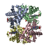
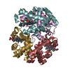
- PDB-1fn3: CRYSTAL STRUCTURE OF NICKEL RECONSTITUTED HEMOGLOBIN-A CASE FOR P... -
+ Open data

- Basic information
| Entry | Database: PDB / ID: 1fn3 | ||||||
|---|---|---|---|---|---|---|---|
| Title | CRYSTAL STRUCTURE OF NICKEL RECONSTITUTED HEMOGLOBIN-A CASE FOR PERMANENT, T-STATE HEMOGLOBIN | ||||||
 Components Components |
| ||||||
 Keywords Keywords | OXYGEN STORAGE/TRANSPORT / PERMANENT T-STATE / METAL ION COORDINATION / SUBUNIT INEQUIVALENCE / SPECTROSCOPY / CRYSTALLOGRAPHY. / OXYGEN STORAGE-TRANSPORT COMPLEX | ||||||
| Function / homology |  Function and homology information Function and homology informationcellular oxidant detoxification / Heme assimilation / nitric oxide transport / hemoglobin alpha binding / hemoglobin binding / haptoglobin-hemoglobin complex / renal absorption / hemoglobin complex / oxygen transport / Scavenging of heme from plasma ...cellular oxidant detoxification / Heme assimilation / nitric oxide transport / hemoglobin alpha binding / hemoglobin binding / haptoglobin-hemoglobin complex / renal absorption / hemoglobin complex / oxygen transport / Scavenging of heme from plasma / erythrocyte development / endocytic vesicle lumen / blood vessel diameter maintenance / hydrogen peroxide catabolic process / oxygen carrier activity / carbon dioxide transport / response to hydrogen peroxide / Heme signaling / Erythrocytes take up oxygen and release carbon dioxide / Erythrocytes take up carbon dioxide and release oxygen / Cytoprotection by HMOX1 / oxygen binding / Late endosomal microautophagy / platelet aggregation / regulation of blood pressure / Chaperone Mediated Autophagy / positive regulation of nitric oxide biosynthetic process / tertiary granule lumen / Factors involved in megakaryocyte development and platelet production / blood microparticle / ficolin-1-rich granule lumen / iron ion binding / inflammatory response / heme binding / Neutrophil degranulation / : / extracellular exosome / extracellular region / membrane / metal ion binding / cytosol Similarity search - Function | ||||||
| Biological species |  Homo sapiens (human) Homo sapiens (human) | ||||||
| Method |  X-RAY DIFFRACTION / MOLECULAR REPLACEMENT / Resolution: 2.48 Å X-RAY DIFFRACTION / MOLECULAR REPLACEMENT / Resolution: 2.48 Å | ||||||
 Authors Authors | Venkateshrao, S. / Deepthi, S. / Pattabhi, V. / Manoharan, P.T. | ||||||
 Citation Citation | Journal: CURR.SCI. / Year: 2003 Title: Crystal Structure of Nickel Reconstituted Hemoglobin - A Case for Permanent, T-State Hemoglobin Authors: Venkateshrao, S. / Deepthi, S. / Pattabhi, V. / Manoharan, P.T. #1: Journal: Protein Sci. / Year: 2000Title: Oxygen Binding by Alpha(Fe2+)2Beta(Ni2+)2 Hemoglobin Crystals Authors: Bruno, S. / Bettati, S. / Manfredini, M. / Mozzarelli, A. / Bolognesi, M. / Deriu, D. / Rosano, C. / Tsuneshige, A. / Yonetani, T. / Henry, E. #2: Journal: J.Mol.Biol. / Year: 1992Title: High Resolution Crystal Structures and Comparisons of T-State Deoxyhaemoglobin and Two Liganded T-State Haemoglobins:T(Alpha-Oxy)Haemoglobin and T(met)Haemoglobin Authors: Liddington, R. / Derewenda, Z. / Dodson, E. / Hubbard, R. / Dodson, G. #3: Journal: J.Mol.Biol. / Year: 1990Title: Structure of Deoxy Quaternary Hemoglobin with Liganded Beta Subunits Authors: Luisi, B. / Liddington, B. / Fermi, G. / Shibayama, N. | ||||||
| History |
|
- Structure visualization
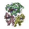
| Structure viewer | Molecule: MolmilJmol/JSmol |
|---|
- Downloads & links
-Download
| PDBx/mmCIF format | 1fn3.cif.gz | 122.9 KB | Display | PDBx/mmCIF format |
|---|---|---|---|---|
| PDB format | pdb1fn3.ent.gz | 97.3 KB | Display | PDB format |
| PDBx/mmJSON format | 1fn3.json.gz | Tree view | PDBx/mmJSON format | |
| Others |  Other downloads Other downloads |
-Validation report
| Arichive directory | https://data.pdbj.org/pub/pdb/validation_reports/fn/1fn3ftp://data.pdbj.org/pub/pdb/validation_reports/fn/1fn3 | HTTPS FTP |
|---|
-Related structure data
| Related structure data | 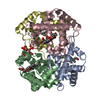 1thbS S: Starting model for refinement |
|---|---|
| Similar structure data |


-Links
 PDBj
PDBj


- Assembly
| Deposited unit | 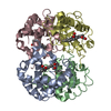
| ||||||||
|---|---|---|---|---|---|---|---|---|---|
| 1 |
| ||||||||
| Unit cell |
|
-Components
| #1: Protein | Mass: 15150.353 Da / Num. of mol.: 2 / Source method: isolated from a natural source / Source: (natural) Homo sapiens (human) / References: UniProt: P69905#2: Protein | Mass: 15890.198 Da / Num. of mol.: 2 / Source method: isolated from a natural source / Source: (natural) Homo sapiens (human) / References: UniProt: P68871#3: Chemical | ChemComp-HNI /   Mass: 619.336 Da / Num. of mol.: 4 / Source method: obtained synthetically / Formula: C34H32N4NiO4 Mass: 619.336 Da / Num. of mol.: 4 / Source method: obtained synthetically / Formula: C34H32N4NiO4#4: Water | ChemComp-HOH / |  Mass: 18.015 Da / Num. of mol.: 38 / Source method: isolated from a natural source / Formula: H2O Mass: 18.015 Da / Num. of mol.: 38 / Source method: isolated from a natural source / Formula: H2O |
|---|
-Experimental details
-Experiment
| Experiment | Method: X-RAY DIFFRACTION / Number of used crystals: 1 |
|---|
- Sample preparation
| Crystal | Density Matthews: 2.41 Å3/Da / Density % sol: 48.95 % | |||||||||||||||
|---|---|---|---|---|---|---|---|---|---|---|---|---|---|---|---|---|
| Crystal grow | pH: 7.2 / Details: pH 7.20 | |||||||||||||||
| Crystal grow | *PLUS Method: batch method | |||||||||||||||
| Components of the solutions | *PLUS
|
-Data collection
| Diffraction | Mean temperature: 293 K |
|---|---|
| Diffraction source | Source: ROTATING ANODE / Wavelength: 1.54 |
| Detector | Type: MARRESEARCH / Detector: IMAGE PLATE / Date: Oct 5, 1999 |
| Radiation | Protocol: SINGLE WAVELENGTH / Monochromatic (M) / Laue (L): M / Scattering type: x-ray |
| Radiation wavelength | Wavelength: 1.54 Å / Relative weight: 1 |
| Reflection | Resolution: 2.48→19.81 Å / Num. obs: 19946 / % possible obs: 91.37 % / Observed criterion σ(I): 0 / Redundancy: 3.8 % / Rmerge(I) obs: 0.1549 / Net I/σ(I): 3.04 |
| Reflection shell | Resolution: 2.48→2.6 Å / Redundancy: 2.9 % / Rmerge(I) obs: 0.7552 / Mean I/σ(I) obs: 0.07 / % possible all: 71.5 |
| Reflection | *PLUS Highest resolution: 2.5 Å / Lowest resolution: 19.8 Å / % possible obs: 91.4 % / Redundancy: 3.8 % / Num. measured all: 75819 / Rmerge(I) obs: 0.155 |
- Processing
| Software |
| |||||||||||||||||||||||||
|---|---|---|---|---|---|---|---|---|---|---|---|---|---|---|---|---|---|---|---|---|---|---|---|---|---|---|
| Refinement | Method to determine structure: MOLECULAR REPLACEMENT Starting model: 1THB Resolution: 2.48→8 Å / Cross valid method: FREE R / σ(F): 2
| |||||||||||||||||||||||||
| Refinement step | Cycle: LAST / Resolution: 2.48→8 Å
| |||||||||||||||||||||||||
| Refine LS restraints |
| |||||||||||||||||||||||||
| Refinement | *PLUS Highest resolution: 2.5 Å | |||||||||||||||||||||||||
| Solvent computation | *PLUS | |||||||||||||||||||||||||
| Displacement parameters | *PLUS | |||||||||||||||||||||||||
| Refine LS restraints | *PLUS
|
