 Movie
Movie Controller

[English] 日本語
 Yorodumi
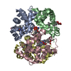
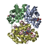
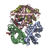
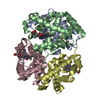
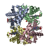
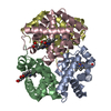
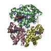
- PDB-1hbs: REFINED CRYSTAL STRUCTURE OF DEOXYHEMOGLOBIN S. I. RESTRAINED LEA... -
+ Open data

- Basic information
| Entry | Database: PDB / ID: 1hbs | ||||||
|---|---|---|---|---|---|---|---|
| Title | REFINED CRYSTAL STRUCTURE OF DEOXYHEMOGLOBIN S. I. RESTRAINED LEAST-SQUARES REFINEMENT AT 3.0-ANGSTROMS RESOLUTION | ||||||
 Components Components |
| ||||||
 Keywords Keywords | OXYGEN TRANSPORT | ||||||
| Function / homology |  Function and homology information Function and homology informationcellular oxidant detoxification / Heme assimilation / nitric oxide transport / hemoglobin alpha binding / hemoglobin binding / haptoglobin-hemoglobin complex / renal absorption / hemoglobin complex / oxygen transport / Scavenging of heme from plasma ...cellular oxidant detoxification / Heme assimilation / nitric oxide transport / hemoglobin alpha binding / hemoglobin binding / haptoglobin-hemoglobin complex / renal absorption / hemoglobin complex / oxygen transport / Scavenging of heme from plasma / erythrocyte development / endocytic vesicle lumen / blood vessel diameter maintenance / hydrogen peroxide catabolic process / oxygen carrier activity / carbon dioxide transport / response to hydrogen peroxide / Heme signaling / Erythrocytes take up oxygen and release carbon dioxide / Erythrocytes take up carbon dioxide and release oxygen / Cytoprotection by HMOX1 / oxygen binding / Late endosomal microautophagy / platelet aggregation / regulation of blood pressure / Chaperone Mediated Autophagy / positive regulation of nitric oxide biosynthetic process / tertiary granule lumen / Factors involved in megakaryocyte development and platelet production / blood microparticle / ficolin-1-rich granule lumen / iron ion binding / inflammatory response / heme binding / Neutrophil degranulation / : / extracellular exosome / extracellular region / membrane / metal ion binding / cytosol Similarity search - Function | ||||||
| Biological species |  Homo sapiens (human) Homo sapiens (human) | ||||||
| Method |  X-RAY DIFFRACTION / Resolution: 3 Å X-RAY DIFFRACTION / Resolution: 3 Å | ||||||
 Authors Authors | Padlan, E.A. / Love, W.E. | ||||||
 Citation Citation | Journal: J.Biol.Chem. / Year: 1985 Title: Refined crystal structure of deoxyhemoglobin S. I. Restrained least-squares refinement at 3.0-A resolution. Authors: Padlan, E.A. / Love, W.E. #1: Journal: J.Biol.Chem. / Year: 1985Title: Refined Crystal Structure of Deoxyhemoglobin S. II. Molecular Interactions in the Crystal Authors: Padlan, E.A. / Love, W.E. #2: Journal: Inserm Symp. / Year: 1978Title: Intermolecular Interactions in Crystals of Human Deoxy Hemoglobin A, C, F and S Authors: Love, W.E. / Fitzgerald, P.M.D. / Hanson, J.C. / Royerjunior, W.E. #3: Journal: MOLECULAR STRUCTURE AND BIOLOGICAL ACTIVITY / Year: 1976Title: Crystal Structure of Sickle-Cell Deoxyhemoglobin Authors: Wishner, B.C. / Hanson, J.C. / Ringle, W.M. / Love, W.E. #4: Journal: J.Mol.Biol. / Year: 1975Title: Crystal Structure of Sickle-Cell Deoxyhemoglobin at 5 Angstroms Resolution Authors: Wishner, B.C. / Ward, K.B. / Lattman, E.E. / Love, W.E. #5: Journal: Proceedings of the First National Symposium on Sickle Cell DiseaseYear: 1975 Title: Crystals of Deoxy Sickle Cell Hemoglobin Authors: Wishner, B.C. / Love, W.E. | ||||||
| History |
|
- Structure visualization
| Structure viewer | Molecule: MolmilJmol/JSmol |
|---|
- Downloads & links
-Download
| PDBx/mmCIF format | 1hbs.cif.gz | 312.6 KB | Display | PDBx/mmCIF format |
|---|---|---|---|---|
| PDB format | pdb1hbs.ent.gz | 184.8 KB | Display | PDB format |
| PDBx/mmJSON format | 1hbs.json.gz | Tree view | PDBx/mmJSON format | |
| Others |  Other downloads Other downloads |
-Validation report
| Arichive directory | https://data.pdbj.org/pub/pdb/validation_reports/hb/1hbsftp://data.pdbj.org/pub/pdb/validation_reports/hb/1hbs | HTTPS FTP |
|---|
-Related structure data
| Similar structure data |
|---|


-Links
 PDBj
PDBj


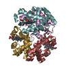
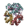
- Assembly
| Deposited unit | 
| ||||||||
|---|---|---|---|---|---|---|---|---|---|
| 1 | 
| ||||||||
| 2 | 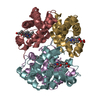
| ||||||||
| Unit cell |
| ||||||||
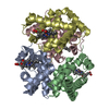
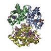
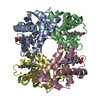
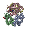
| Noncrystallographic symmetry (NCS) | NCS oper: (Code: given Matrix: (0.9997, -0.0211, 0.01274), Vector: Details | THERE ARE TWO DEOXY HEMOGLOBIN TETRAMERS IN THE ASYMMETRIC UNIT. CHAINS A, B, C, D CONSTITUTE THE FIRST TETRAMER AND CHAINS E, F, G, H CONSTITUTE THE SECOND. THESE TETRAMERS ARE RELATED APPROXIMATELY BY A NON-CRYSTALLOGRAPHIC TWO-FOLD SCREW AXIS PARALLEL TO THE A-AXIS OF THE CRYSTAL. APPLYING THE TRANSFORMATION GIVEN IN THE *MTRIX* RECORDS BELOW TO THE SECOND TETRAMER WILL YIELD APPROXIMATE COORDINATES FOR THE FIRST TETRAMER. | |
-Components
| #1: Protein | Mass: 15150.353 Da / Num. of mol.: 4 Source method: isolated from a genetically manipulated source Source: (gene. exp.) Homo sapiens (human) / References: UniProt: P69905#2: Protein | Mass: 15860.216 Da / Num. of mol.: 4 Source method: isolated from a genetically manipulated source Source: (gene. exp.) Homo sapiens (human) / References: UniProt: P68871#3: Chemical | ChemComp-HEM /   Mass: 616.487 Da / Num. of mol.: 8 / Source method: obtained synthetically / Formula: C34H32FeN4O4 Mass: 616.487 Da / Num. of mol.: 8 / Source method: obtained synthetically / Formula: C34H32FeN4O4 |
|---|
-Experimental details
-Experiment
| Experiment | Method: X-RAY DIFFRACTION |
|---|
- Sample preparation
| Crystal | Density Matthews: 2.51 Å3/Da / Density % sol: 50.93 % |
|---|---|
| Crystal grow | *PLUS Method: unknown / PH range low: 5 / PH range high: 4 |
| Components of the solutions | *PLUS Common name: PEG6000 |
-Data collection
| Radiation | Scattering type: x-ray |
|---|---|
| Radiation wavelength | Relative weight: 1 |
| Reflection | *PLUS Highest resolution: 3 Å / Lowest resolution: 10 Å / Num. obs: 22633 / Rmerge(I) obs: 0.382 |
- Processing
| Refinement | Rfactor obs: 0.254 / Highest resolution: 3 Å | ||||||||||||
|---|---|---|---|---|---|---|---|---|---|---|---|---|---|
| Refinement step | Cycle: LAST / Highest resolution: 3 Å
| ||||||||||||
| Refinement | *PLUS Lowest resolution: 5 Å / Num. reflection obs: 17662 | ||||||||||||
| Solvent computation | *PLUS | ||||||||||||
| Displacement parameters | *PLUS |
