Resolution: 2→10 Å / Isotropic thermal model: ISOTROPIC / Cross valid method: THROUGHOUT / σ(F): 0 / σ(I): 0 / Details: Average Isotropic B Value for Main Chain Atoms.
Rfactor
Num. reflection
% reflection
Selection details
Rfree
0.283
2471
-
RANDOM
Rwork
0.225
-
-
-
all
-
26661
-
-
obs
-
24846
93.2 %
-
Displacement parameters
Biso mean: 27.783 Å2
Refine analyze
Luzzati sigma a obs: 0.328 Å
Refinement step
Cycle: LAST / Resolution: 2→10 Å
Protein
Nucleic acid
Ligand
Solvent
Total
Num. atoms
4384
0
216
220
4820
Refine LS restraints
Refine-ID
Type
Dev ideal
X-RAY DIFFRACTION
x_bond_d
0.014
X-RAY DIFFRACTION
x_angle_deg
1.813
+
About Yorodumi
-
News
-
Feb 9, 2022. New format data for meta-information of EMDB entries
New format data for meta-information of EMDB entries
Version 3 of the EMDB header file is now the official format.
The previous official version 1.9 will be removed from the archive.
In the structure databanks used in Yorodumi, some data are registered as the other names, "COVID-19 virus" and "2019-nCoV". Here are the details of the virus and the list of structure data.
Jan 31, 2019. EMDB accession codes are about to change! (news from PDBe EMDB page)
EMDB accession codes are about to change! (news from PDBe EMDB page)
The allocation of 4 digits for EMDB accession codes will soon come to an end. Whilst these codes will remain in use, new EMDB accession codes will include an additional digit and will expand incrementally as the available range of codes is exhausted. The current 4-digit format prefixed with “EMD-” (i.e. EMD-XXXX) will advance to a 5-digit format (i.e. EMD-XXXXX), and so on. It is currently estimated that the 4-digit codes will be depleted around Spring 2019, at which point the 5-digit format will come into force.
The EM Navigator/Yorodumi systems omit the EMD- prefix.
Related info.:Q: What is EMD? / ID/Accession-code notation in Yorodumi/EM Navigator
Yorodumi is a browser for structure data from EMDB, PDB, SASBDB, etc.
This page is also the successor to EM Navigator detail page, and also detail information page/front-end page for Omokage search.
The word "yorodu" (or yorozu) is an old Japanese word meaning "ten thousand". "mi" (miru) is to see.
Related info.:EMDB / PDB / SASBDB / Comparison of 3 databanks / Yorodumi Search / Aug 31, 2016. New EM Navigator & Yorodumi / Yorodumi Papers / Jmol/JSmol / Function and homology information / Changes in new EM Navigator and Yorodumi
 Movie
Movie Controller

 Yorodumi
 Open data
 Basic information
 Components
 Keywords
 Function and homology information
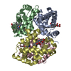
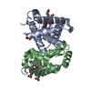
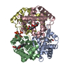
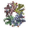
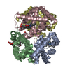
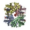
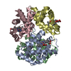
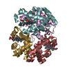
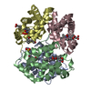
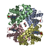
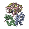
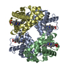
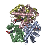
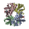
 Homo sapiens (human)
 X-RAY DIFFRACTION /
  Authors
 Citation
 Structure visualization
 Downloads & links
 Other downloads


 PDBj
PDBj


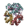
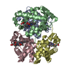
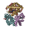
 Assembly


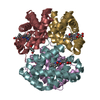
 Mass: 616.487 Da / Num. of mol.: 4 / Source method: obtained synthetically / Formula: C34H32FeN4O4
 Mass: 28.010 Da / Num. of mol.: 4 / Source method: obtained synthetically / Formula: CO
 Mass: 660.035 Da / Num. of mol.: 1 / Source method: obtained synthetically / Formula: C6H18O24P6
 Sample preparation
 / Beamline: D03B-MX1 / Wavelength: 1.54 Å
 Processing