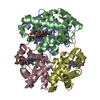
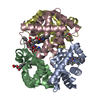
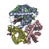
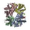
Resolution: 2.4→30 Å / σ(F): 0 Details: Author provided the following evidence of direct S1P binding to hemoglobin: S1P beads successfully pulled down HbA from normal human erythrocyte lysates, while sphingosine or ...Details: Author provided the following evidence of direct S1P binding to hemoglobin: S1P beads successfully pulled down HbA from normal human erythrocyte lysates, while sphingosine or lysophosphatidic acid beads failed to pull down Hb. In addition, co-binding of 2,3-DPG and S1P to hemoglobin (Hb) is required for S1P-induced decrease in Hb-O2 affinity. We measured Oxygen Equilibration Curves (OEC) of purified HbA in the presence of 2,3-DPG along with S1P. We found that S1P decreased HbA-O2 binding affinity with increased P50 in a dose-dependent manner. Thus, we provide biochemical and functional evidence that S1P binds directly to Hb but requires cobinding of 2,3-DPG to decrease the protein affinity for O2
In the structure databanks used in Yorodumi, some data are registered as the other names, "COVID-19 virus" and "2019-nCoV". Here are the details of the virus and the list of structure data.
Jan 31, 2019. EMDB accession codes are about to change! (news from PDBe EMDB page)
EMDB accession codes are about to change! (news from PDBe EMDB page)
The allocation of 4 digits for EMDB accession codes will soon come to an end. Whilst these codes will remain in use, new EMDB accession codes will include an additional digit and will expand incrementally as the available range of codes is exhausted. The current 4-digit format prefixed with “EMD-” (i.e. EMD-XXXX) will advance to a 5-digit format (i.e. EMD-XXXXX), and so on. It is currently estimated that the 4-digit codes will be depleted around Spring 2019, at which point the 5-digit format will come into force.
The EM Navigator/Yorodumi systems omit the EMD- prefix.
Related info.:Q: What is EMD? / ID/Accession-code notation in Yorodumi/EM Navigator
Yorodumi is a browser for structure data from EMDB, PDB, SASBDB, etc.
This page is also the successor to EM Navigator detail page, and also detail information page/front-end page for Omokage search.
The word "yorodu" (or yorozu) is an old Japanese word meaning "ten thousand". "mi" (miru) is to see.
Related info.:EMDB / PDB / SASBDB / Comparison of 3 databanks / Yorodumi Search / Aug 31, 2016. New EM Navigator & Yorodumi / Yorodumi Papers / Jmol/JSmol / Function and homology information / Changes in new EM Navigator and Yorodumi
 Movie
Movie Controller

 Yorodumi
 Open data
 Basic information
 Components
 Keywords
 Function and homology information
 Homo sapiens (human)
 X-RAY DIFFRACTION /
  Authors
 United States, 2items
  Citation
 Structure visualization
 Downloads & links
 Other downloads


 PDBj
PDBj


 Assembly

 Mass: 616.487 Da / Num. of mol.: 4 / Source method: obtained synthetically / Formula: C34H32FeN4O4
 Mass: 379.472 Da / Num. of mol.: 2 / Source method: obtained synthetically / Formula: C18H38NO5P
 Mass: 18.015 Da / Num. of mol.: 545 / Source method: isolated from a natural source / Formula: H2O
 Sample preparation
 Processing