 Movie
Movie Controller


+ Open data

- Basic information
| Entry | Database: PDB / ID: 1ol0 | ||||||
|---|---|---|---|---|---|---|---|
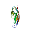
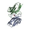
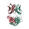
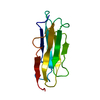
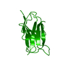
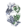
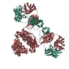
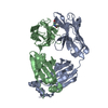
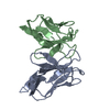
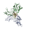
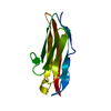
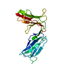
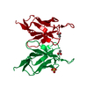
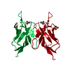
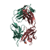
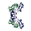
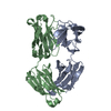
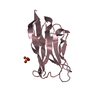
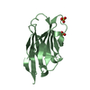
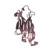
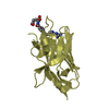
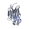
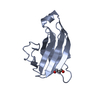
| Title | Crystal structure of a camelised human VH | ||||||
 Components Components | IMMUNOGLOBULIN G | ||||||
 Keywords Keywords | IMMUNOGLOBULIN / CAMELISED VARIABLE HEAVY DOMAIN | ||||||
| Function / homology | Immunoglobulins / Immunoglobulin-like / Sandwich / Mainly Beta Function and homology information Function and homology information | ||||||
| Biological species |  HOMO SAPIENS (human) HOMO SAPIENS (human) | ||||||
| Method |  X-RAY DIFFRACTION / SYNCHROTRON / MOLECULAR REPLACEMENT / Resolution: 1.8 Å X-RAY DIFFRACTION / SYNCHROTRON / MOLECULAR REPLACEMENT / Resolution: 1.8 Å | ||||||
 Authors Authors | Dottorini, T. / Vaughan, C.K. / Walsh, M.A. / Losurdo, P. / Sollazzo, M. | ||||||
 Citation Citation | Journal: Biochemistry / Year: 2004 Title: Crystal Structure of a Human Vh: Requirements for Maintaining a Monomeric Fragment Authors: Dottorini, T. / Vaughan, C.K. / Walsh, M.A. / Losurdo, P. / Sollazzo, M. #1: Journal: J.Biol.Chem. / Year: 2000Title: Inhibition of the Hepatitis C Virus Ns3/4A Protease. The Crystal Structures of Two Protease-Inhibitor Complexes Authors: Dimarco, S. / Rizzi, M. / Volpari, C. / Walsh, M.A. / Narjes, F. / Colarusso, S. / Defrancesco, R. / Matassa, V.G. / Sollazzo, M. #2: Journal: Protein Eng. / Year: 1997 Title: Affinity Selection of a Camelized V(H) Domain Antibody Inhibitor of Hepatitis C Virus Ns3 Protease Authors: Martin, F. / Volpari, C. / Steinkuhler, C. / Dimasibrunetti, M. / Biasiol, G. / Altamura, S. / Cortese, R. / Defrancesco, R. / Sollazzo, M. #3: Journal: J.Mol.Biol. / Year: 1996Title: Rearrangement of the Former Vl Interface in the Solution Structure of a Camelised, Single Antibody Vh Domain Authors: Riechmann, L. | ||||||
| History |
| ||||||
| Remark 700 | SHEET THE SHEET STRUCTURE OF THIS MOLECULE IS BIFURCATED. IN ORDER TO REPRESENT THIS FEATURE IN ... SHEET THE SHEET STRUCTURE OF THIS MOLECULE IS BIFURCATED. IN ORDER TO REPRESENT THIS FEATURE IN THE SHEET RECORDS BELOW, TWO SHEETS ARE DEFINED. |
- Structure visualization
| Structure viewer | Molecule: MolmilJmol/JSmol |
|---|
- Downloads & links
-Download
| PDBx/mmCIF format | 1ol0.cif.gz | 70.4 KB | Display | PDBx/mmCIF format |
|---|---|---|---|---|
| PDB format | pdb1ol0.ent.gz | 53.8 KB | Display | PDB format |
| PDBx/mmJSON format | 1ol0.json.gz | Tree view | PDBx/mmJSON format | |
| Others |  Other downloads Other downloads |
-Validation report
| Arichive directory | https://data.pdbj.org/pub/pdb/validation_reports/ol/1ol0ftp://data.pdbj.org/pub/pdb/validation_reports/ol/1ol0 | HTTPS FTP |
|---|
-Related structure data
| Related structure data |  1hcvS S: Starting model for refinement |
|---|---|
| Similar structure data |


-Links
 PDBj
PDBj

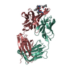
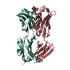
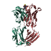
- Assembly
| Deposited unit | 
| ||||||||
|---|---|---|---|---|---|---|---|---|---|
| 1 | 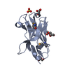
| ||||||||
| 2 | 
| ||||||||
| Unit cell |
| ||||||||
| Noncrystallographic symmetry (NCS) | NCS oper: (Code: given Matrix: (-0.999943, -0.009568, -0.00477), Vector: |
-Components
| #1: Antibody | Mass: 13254.387 Da / Num. of mol.: 2 / Fragment: VARIABLE HEAVY DOMAIN Source method: isolated from a genetically manipulated source Source: (gene. exp.) HOMO SAPIENS (human) / Production host:  #2: Chemical | ChemComp-SO4 /   Mass: 96.063 Da / Num. of mol.: 5 / Source method: obtained synthetically / Formula: SO4 Mass: 96.063 Da / Num. of mol.: 5 / Source method: obtained synthetically / Formula: SO4#3: Chemical | ChemComp-GOL /   Mass: 92.094 Da / Num. of mol.: 5 / Source method: obtained synthetically / Formula: C3H8O3 Mass: 92.094 Da / Num. of mol.: 5 / Source method: obtained synthetically / Formula: C3H8O3#4: Water | ChemComp-HOH / |  Mass: 18.015 Da / Num. of mol.: 388 / Source method: isolated from a natural source / Formula: H2O Mass: 18.015 Da / Num. of mol.: 388 / Source method: isolated from a natural source / Formula: H2OHas protein modification | Y | |
|---|
-Experimental details
-Experiment
| Experiment | Method: X-RAY DIFFRACTION / Number of used crystals: 1 |
|---|
- Sample preparation
| Crystal | Density Matthews: 3.1 Å3/Da / Density % sol: 59.5 % | ||||||||||||||||||||||||||||||
|---|---|---|---|---|---|---|---|---|---|---|---|---|---|---|---|---|---|---|---|---|---|---|---|---|---|---|---|---|---|---|---|
| Crystal grow | Method: vapor diffusion, hanging drop / pH: 7.5 Details: CRYSTALS GREW FROM 4 MICROLITRE HANGING DROPS FORMED BY MIXING 2 MICROLITERS OF PROTEIN AT A CONCENTRATION OF 5MG/ML WITH 2 MICROLITERS OF RESERVOIR SOLUTION CONTAINING 15-20% (W/V) PEG ...Details: CRYSTALS GREW FROM 4 MICROLITRE HANGING DROPS FORMED BY MIXING 2 MICROLITERS OF PROTEIN AT A CONCENTRATION OF 5MG/ML WITH 2 MICROLITERS OF RESERVOIR SOLUTION CONTAINING 15-20% (W/V) PEG 8000, 0.4 M AMMONIUM SULPHATE AND 0.1M HEPES AT PH 7.5. | ||||||||||||||||||||||||||||||
| Crystal grow | *PLUS pH: 7.5 / Method: vapor diffusion, hanging drop | ||||||||||||||||||||||||||||||
| Components of the solutions | *PLUS
|
-Data collection
| Diffraction | Mean temperature: 100 K |
|---|---|
| Diffraction source | Source: SYNCHROTRON / Site: ESRF  / Beamline: ID14-2 / Wavelength: 0.933 / Beamline: ID14-2 / Wavelength: 0.933 |
| Detector | Type: ADSC CCD / Detector: CCD / Date: Oct 15, 2000 / Details: TOROIDAL |
| Radiation | Monochromator: DIAMON 100/GE 220 / Protocol: SINGLE WAVELENGTH / Monochromatic (M) / Laue (L): M / Scattering type: x-ray |
| Radiation wavelength | Wavelength: 0.933 Å / Relative weight: 1 |
| Reflection | Resolution: 1.8→30 Å / Num. obs: 29910 / % possible obs: 94.9 % / Observed criterion σ(I): -3 / Redundancy: 2.3 % / Rmerge(I) obs: 0.051 / Net I/σ(I): 17.2 |
| Reflection shell | Resolution: 1.8→1.83 Å / Redundancy: 2 % / Rmerge(I) obs: 0.227 / Mean I/σ(I) obs: 1.5 / % possible all: 71.5 |
| Reflection | *PLUS Highest resolution: 1.8 Å / Lowest resolution: 25.2 Å / Num. measured all: 69437 / Rmerge(I) obs: 0.051 |
| Reflection shell | *PLUS Lowest resolution: 1.84 Å / % possible obs: 71.5 % / Rmerge(I) obs: 0.227 / Mean I/σ(I) obs: 1.5 |
- Processing
| Software |
| ||||||||||||||||||||
|---|---|---|---|---|---|---|---|---|---|---|---|---|---|---|---|---|---|---|---|---|---|
| Refinement | Method to determine structure: MOLECULAR REPLACEMENT Starting model: PDB ENTRY 1HCV Resolution: 1.8→25.24 Å / SU B: 1.401 / SU ML: 0.044 / Cross valid method: THROUGHOUT EXCEPT IN LAST RO / ESU R: 0.095
| ||||||||||||||||||||
| Displacement parameters | Biso mean: 17.342 Å2
| ||||||||||||||||||||
| Refinement step | Cycle: LAST / Resolution: 1.8→25.24 Å
| ||||||||||||||||||||
| Refinement | *PLUS Highest resolution: 1.8 Å / Lowest resolution: 25.2 Å / % reflection Rfree: 5 % / Rfactor Rfree: 0.1802 / Rfactor Rwork: 0.1558 | ||||||||||||||||||||
| Solvent computation | *PLUS | ||||||||||||||||||||
| Displacement parameters | *PLUS | ||||||||||||||||||||
| Refine LS restraints | *PLUS
|
