 Movie
Movie Controller

[English] 日本語
 Yorodumi
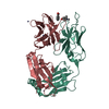
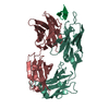
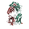
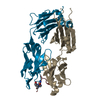
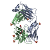
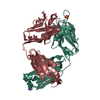
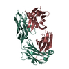
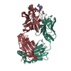
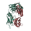
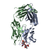
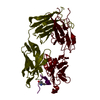
- PDB-1aj7: IMMUNOGLOBULIN 48G7 GERMLINE FAB ANTIBODY COMPLEXED WITH HAPTEN 5... -
+ Open data

- Basic information
| Entry | Database: PDB / ID: 1aj7 | ||||||
|---|---|---|---|---|---|---|---|
| Title | IMMUNOGLOBULIN 48G7 GERMLINE FAB ANTIBODY COMPLEXED WITH HAPTEN 5-(PARA-NITROPHENYL PHOSPHONATE)-PENTANOIC ACID. AFFINITY MATURATION OF AN ESTEROLYTIC ANTIBODY | ||||||
 Components Components |
| ||||||
 Keywords Keywords | IMMUNOGLOBULIN / GERMLINE ANTIBODY / FAB / CATALYTIC ANTIBODY / AFFINITY MATURATION | ||||||
| Function / homology |  Function and homology information Function and homology informationcomplement-dependent cytotoxicity / antibody-dependent cellular cytotoxicity / Fc-gamma receptor I complex binding / immunoglobulin complex, circulating / Classical antibody-mediated complement activation / immunoglobulin receptor binding / IgG immunoglobulin complex / Initial triggering of complement / FCGR activation / complement activation, classical pathway ...complement-dependent cytotoxicity / antibody-dependent cellular cytotoxicity / Fc-gamma receptor I complex binding / immunoglobulin complex, circulating / Classical antibody-mediated complement activation / immunoglobulin receptor binding / IgG immunoglobulin complex / Initial triggering of complement / FCGR activation / complement activation, classical pathway / Role of phospholipids in phagocytosis / antigen binding / FCGR3A-mediated IL10 synthesis / Regulation of Complement cascade / B cell receptor signaling pathway / FCGR3A-mediated phagocytosis / Regulation of actin dynamics for phagocytic cup formation / antibacterial humoral response / Interleukin-4 and Interleukin-13 signaling / blood microparticle / adaptive immune response / : / extracellular exosome / extracellular region / plasma membrane Similarity search - Function | ||||||
| Biological species |   Homo sapiens (human) Homo sapiens (human) | ||||||
| Method |  X-RAY DIFFRACTION / SYNCHROTRON / MOLECULAR REPLACEMENT / Resolution: 2.1 Å X-RAY DIFFRACTION / SYNCHROTRON / MOLECULAR REPLACEMENT / Resolution: 2.1 Å | ||||||
 Authors Authors | Wedemayer, G.J. / Wang, L.H. / Patten, P.A. / Schultz, P.G. / Stevens, R.C. | ||||||
 Citation Citation | Journal: Science / Year: 1997 Title: Structural insights into the evolution of an antibody combining site. Authors: Wedemayer, G.J. / Patten, P.A. / Wang, L.H. / Schultz, P.G. / Stevens, R.C. #1: Journal: J.Mol.Biol. / Year: 1997Title: Crystal Structures of the Free and Liganded Form of an Esterolytic Catalytic Antibody Authors: Wedemayer, G.J. / Wang, L.H. / Patten, P.A. / Schultz, P.G. / Stevens, R.C. #2: Journal: Science / Year: 1996Title: The Immunological Evolution of Catalysis Authors: Patten, P.A. / Gray, N.S. / Yang, P.L. / Marks, C.B. / Wedemayer, G.J. / Boniface, J.J. / Stevens, R.C. / Schultz, P.G. #3: Journal: Proc.Natl.Acad.Sci.USA / Year: 1993Title: A Genetic Approach to the Generation of Antibodies with Enhanced Catalytic Activities Authors: Lesley, S.A. / Patten, P.A. / Schultz, P.G. | ||||||
| History |
|
- Structure visualization
| Structure viewer | Molecule: MolmilJmol/JSmol |
|---|
- Downloads & links
-Download
| PDBx/mmCIF format | 1aj7.cif.gz | 110.5 KB | Display | PDBx/mmCIF format |
|---|---|---|---|---|
| PDB format | pdb1aj7.ent.gz | 84.7 KB | Display | PDB format |
| PDBx/mmJSON format | 1aj7.json.gz | Tree view | PDBx/mmJSON format | |
| Others |  Other downloads Other downloads |
-Validation report
| Arichive directory | https://data.pdbj.org/pub/pdb/validation_reports/aj/1aj7ftp://data.pdbj.org/pub/pdb/validation_reports/aj/1aj7 | HTTPS FTP |
|---|
-Related structure data
| Related structure data |  2rcsC  1gafS S: Starting model for refinement C: citing same article ( |
|---|---|
| Similar structure data |


-Links
 PDBj
PDBj


- Assembly
| Deposited unit | 
| ||||||||
|---|---|---|---|---|---|---|---|---|---|
| 1 |
| ||||||||
| Unit cell |
|
-Components
| #1: Antibody | Mass: 23464.988 Da / Num. of mol.: 1 Fragment: VARIABLE DOMAINS OF LIGHT AND HEAVY CHAINS AND CONSTANT DOMAINS OF LIGHT AND HEAVY CHAINS Source method: isolated from a genetically manipulated source Source: (gene. exp.) Description: EACH CHAIN IS A FUSION POLYPEPTIDE WHICH IS PART HUMAN AND PART MOUSE Cell line: 48G7 / Fragment: CONSTANT DOMAINS OF LIGHT AND HEAVY CHAINS / Plasmid: PSAL143 / Species (production host): Escherichia coli / Production host:  |
|---|---|
| #2: Antibody | Mass: 23062.750 Da / Num. of mol.: 1 Fragment: VARIABLE DOMAINS OF LIGHT AND HEAVY CHAINS AND CONSTANT DOMAINS OF LIGHT AND HEAVY CHAINS Source method: isolated from a genetically manipulated source Source: (gene. exp.) Homo sapiens (human)Description: EACH CHAIN IS A FUSION POLYPEPTIDE WHICH IS PART HUMAN AND PART MOUSE Cell line: 48G7 / Fragment: CONSTANT DOMAINS OF LIGHT AND HEAVY CHAINS / Plasmid: PSAL143 / Species (production host): Escherichia coli / Production host: |
| #3: Chemical | ChemComp-NPE /   Mass: 302.197 Da / Num. of mol.: 1 / Source method: obtained synthetically / Formula: C11H13NO7P Mass: 302.197 Da / Num. of mol.: 1 / Source method: obtained synthetically / Formula: C11H13NO7P |
| Has protein modification | Y |
-Experimental details
-Experiment
| Experiment | Method: X-RAY DIFFRACTION / Number of used crystals: 1 |
|---|
- Sample preparation
| Crystal | Density Matthews: 2.13 Å3/Da / Density % sol: 42.29 % | ||||||||||||||||||||||||||||||||||||||||||||||||||||||||||||||||||
|---|---|---|---|---|---|---|---|---|---|---|---|---|---|---|---|---|---|---|---|---|---|---|---|---|---|---|---|---|---|---|---|---|---|---|---|---|---|---|---|---|---|---|---|---|---|---|---|---|---|---|---|---|---|---|---|---|---|---|---|---|---|---|---|---|---|---|---|
| Crystal grow | pH: 8 Details: 10-15 MG/ML FAB IN 100MM NACL, 10MM TRIS PH 8.0, 1MM METHIONINE, 1MM SODIUM AZIDE, 0.5 MM EDTA. | ||||||||||||||||||||||||||||||||||||||||||||||||||||||||||||||||||
| Crystal grow | *PLUS Method: vapor diffusion, hanging drop | ||||||||||||||||||||||||||||||||||||||||||||||||||||||||||||||||||
| Components of the solutions | *PLUS
|
-Data collection
| Diffraction | Mean temperature: 100 K |
|---|---|
| Diffraction source | Source: SYNCHROTRON / Site: SSRL  / Beamline: BL7-1 / Wavelength: 1.08 / Beamline: BL7-1 / Wavelength: 1.08 |
| Detector | Type: MARRESEARCH / Detector: IMAGE PLATE / Date: May 1, 1996 |
| Radiation | Monochromatic (M) / Laue (L): M / Scattering type: x-ray |
| Radiation wavelength | Wavelength: 1.08 Å / Relative weight: 1 |
| Reflection | Resolution: 2.1→20 Å / Num. obs: 23005 / % possible obs: 99.5 % / Redundancy: 3.5 % / Rmerge(I) obs: 0.069 / Rsym value: 0.069 / Net I/σ(I): 15 |
| Reflection shell | Resolution: 2.1→2.17 Å / Redundancy: 3.5 % / Rmerge(I) obs: 0.204 / Mean I/σ(I) obs: 5.2 / Rsym value: 0.204 / % possible all: 98.1 |
| Reflection | *PLUS Num. measured all: 80267 |
| Reflection shell | *PLUS % possible obs: 98.1 % |
- Processing
| Software |
| ||||||||||||||||||||||||||||||||||||||||||||||||||||||||||||
|---|---|---|---|---|---|---|---|---|---|---|---|---|---|---|---|---|---|---|---|---|---|---|---|---|---|---|---|---|---|---|---|---|---|---|---|---|---|---|---|---|---|---|---|---|---|---|---|---|---|---|---|---|---|---|---|---|---|---|---|---|---|
| Refinement | Method to determine structure: MOLECULAR REPLACEMENT Starting model: PDB ENTRY 1GAF Resolution: 2.1→20 Å / σ(F): 2
| ||||||||||||||||||||||||||||||||||||||||||||||||||||||||||||
| Refinement step | Cycle: LAST / Resolution: 2.1→20 Å
| ||||||||||||||||||||||||||||||||||||||||||||||||||||||||||||
| Refine LS restraints |
| ||||||||||||||||||||||||||||||||||||||||||||||||||||||||||||
| LS refinement shell | Resolution: 2.1→2.11 Å / Total num. of bins used: 60
|
