 Movie
Movie Controller

[English] 日本語
 Yorodumi
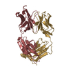
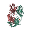
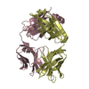
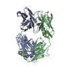
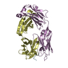
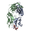
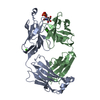
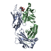
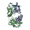
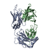
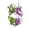
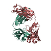
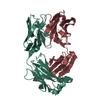
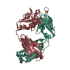
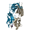
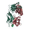
- PDB-1iqw: CRYSTAL STRUCTURE OF THE FAB FRAGMENT OF THE MOUSE ANTI-HUMAN FAS... -
+ Open data

- Basic information
| Entry | Database: PDB / ID: 1iqw | ||||||
|---|---|---|---|---|---|---|---|
| Title | CRYSTAL STRUCTURE OF THE FAB FRAGMENT OF THE MOUSE ANTI-HUMAN FAS ANTIBODY HFE7A | ||||||
 Components Components |
| ||||||
 Keywords Keywords | IMMUNE SYSTEM / IMMUNOGLOBULIN / FAB / ANTI_FAS / AGONISTIC ANTIBODY / APOPTOSIS | ||||||
| Function / homology | Immunoglobulins / Immunoglobulin-like / Sandwich / Mainly Beta Function and homology information Function and homology information | ||||||
| Biological species |  | ||||||
| Method |  X-RAY DIFFRACTION / SYNCHROTRON / MOLECULAR REPLACEMENT / Resolution: 2.5 Å X-RAY DIFFRACTION / SYNCHROTRON / MOLECULAR REPLACEMENT / Resolution: 2.5 Å | ||||||
 Authors Authors | Ito, S. / Takayama, T. / Hanzawa, H. / Ichikawa, K. / Ohsumi, J. / Serizawa, N. / Hata, T. / Haruyama, H. | ||||||
 Citation Citation | Journal: J.Biochem. / Year: 2002 Title: Crystal structure of the antigen-binding fragment of apoptosis-inducing mouse anti-human Fas monoclonal antibody HFE7A. Authors: Ito, S. / Takayama, T. / Hanzawa, H. / Ichikawa, K. / Ohsumi, J. / Serizawa, N. / Hata, T. / Haruyama, H. #1: Journal: Acta Crystallogr.,Sect.D / Year: 2001Title: Crystallization and preliminary X-ray crystallographic studies on a Fab fragment of the mouse anti-human Fas monoclonal antibody HFE7A Authors: Ito, S. / Takayama, T. / Hanzawa, H. / Ichikawa, K. / Ohsumi, J. / Serizawa, N. / Haruyama, H. / Hata, T. #2: Journal: BIOSCI.BIOTECHNOL.BIOCHEM. / Year: 2000Title: Cloning and expression of a novel murine anti-human Fas antibody Authors: Yoshida-Kato, H. / Ichikawa, K. / Yamaguchi, J. / Watanabe, K. / Ohsumi, J. / Yonehara, S. / Serizawa, N. #3: Journal: INT.IMMUNOL. / Year: 2000Title: A novel murine anti-human Fas mAb which mitigates lymphadenopathy without hepatotoxicity Authors: Ichikawa, K. / Yoshida-Kato, H. / Ohtsuki, M. / Ohsumi, J. / Yamaguchi, J. / Takahashi, S. / Tani, Y. / Watanabe, M. / Shiraishi, A. / Nishioka, K. / Yonehara, S. / Serizawa, N. | ||||||
| History |
|
- Structure visualization
| Structure viewer | Molecule: MolmilJmol/JSmol |
|---|
- Downloads & links
-Download
| PDBx/mmCIF format | 1iqw.cif.gz | 95.9 KB | Display | PDBx/mmCIF format |
|---|---|---|---|---|
| PDB format | pdb1iqw.ent.gz | 73.2 KB | Display | PDB format |
| PDBx/mmJSON format | 1iqw.json.gz | Tree view | PDBx/mmJSON format | |
| Others |  Other downloads Other downloads |
-Validation report
| Arichive directory | https://data.pdbj.org/pub/pdb/validation_reports/iq/1iqwftp://data.pdbj.org/pub/pdb/validation_reports/iq/1iqw | HTTPS FTP |
|---|
-Related structure data
| Similar structure data |
|---|


-Links
 PDBj
PDBj

- Assembly
| Deposited unit | 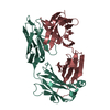
| ||||||||
|---|---|---|---|---|---|---|---|---|---|
| 1 |
| ||||||||
| Unit cell |
|
-Components
| #1: Antibody | Mass: 23919.178 Da / Num. of mol.: 1 / Fragment: FAB FRAGMENT / Source method: isolated from a natural source / Source: (natural) |
|---|---|
| #2: Antibody | Mass: 24886.852 Da / Num. of mol.: 1 / Fragment: FAB FRAGMENT / Source method: isolated from a natural source / Source: (natural) |
| #3: Water | ChemComp-HOH /  Mass: 18.015 Da / Num. of mol.: 57 / Source method: isolated from a natural source / Formula: H2O Mass: 18.015 Da / Num. of mol.: 57 / Source method: isolated from a natural source / Formula: H2O |
| Has protein modification | Y |
-Experimental details
-Experiment
| Experiment | Method: X-RAY DIFFRACTION / Number of used crystals: 1 |
|---|
- Sample preparation
| Crystal | Density Matthews: 2.2 Å3/Da / Density % sol: 44.1 % | ||||||||||||||||||||||||||||||||||||
|---|---|---|---|---|---|---|---|---|---|---|---|---|---|---|---|---|---|---|---|---|---|---|---|---|---|---|---|---|---|---|---|---|---|---|---|---|---|
| Crystal grow | Temperature: 296 K / Method: vapor diffusion, hanging drop / pH: 8.5 Details: SODIUM CITRATE, SODIUM BORATE, pH 8.50, VAPOR DIFFUSION, HANGING DROP, temperature 296.0K | ||||||||||||||||||||||||||||||||||||
| Crystal grow | *PLUS Temperature: 296 K / pH: 7.4 / Details: used seeding | ||||||||||||||||||||||||||||||||||||
| Components of the solutions | *PLUS
|
-Data collection
| Diffraction | Mean temperature: 296 K |
|---|---|
| Diffraction source | Source: SYNCHROTRON / Site: Photon Factory  / Beamline: BL-6B / Wavelength: 1 Å / Beamline: BL-6B / Wavelength: 1 Å |
| Detector | Type: WEISSENBERG / Detector: DIFFRACTOMETER / Date: Dec 5, 1998 |
| Radiation | Protocol: SINGLE WAVELENGTH / Monochromatic (M) / Laue (L): M / Scattering type: x-ray |
| Radiation wavelength | Wavelength: 1 Å / Relative weight: 1 |
| Reflection | Resolution: 2.5→100 Å / Num. obs: 15256 / % possible obs: 97.8 % / Observed criterion σ(I): -3 / Redundancy: 4.6 % / Biso Wilson estimate: 23.7 Å2 / Rmerge(I) obs: 0.045 / Net I/σ(I): 32.3 |
| Reflection shell | Resolution: 2.5→2.59 Å / Rmerge(I) obs: 0.092 / Mean I/σ(I) obs: 10.5 / % possible all: 97.8 |
| Reflection | *PLUS Num. measured all: 69732 |
| Reflection shell | *PLUS % possible obs: 97.8 % |
- Processing
| Software |
| ||||||||||||||||||||||||||||||||||||||||||||||||||||||||||||||||||||||||||||||||
|---|---|---|---|---|---|---|---|---|---|---|---|---|---|---|---|---|---|---|---|---|---|---|---|---|---|---|---|---|---|---|---|---|---|---|---|---|---|---|---|---|---|---|---|---|---|---|---|---|---|---|---|---|---|---|---|---|---|---|---|---|---|---|---|---|---|---|---|---|---|---|---|---|---|---|---|---|---|---|---|---|---|
| Refinement | Method to determine structure: MOLECULAR REPLACEMENT / Resolution: 2.5→40 Å / Rfactor Rfree error: 0.007 / Data cutoff high absF: 10000000 / Data cutoff low absF: 0.001 / Isotropic thermal model: RESTRAINED / Cross valid method: THROUGHOUT / σ(F): 2 / Details: BULK SOLVENT MODEL USED
| ||||||||||||||||||||||||||||||||||||||||||||||||||||||||||||||||||||||||||||||||
| Displacement parameters | Biso mean: 24.5 Å2
| ||||||||||||||||||||||||||||||||||||||||||||||||||||||||||||||||||||||||||||||||
| Refine analyze |
| ||||||||||||||||||||||||||||||||||||||||||||||||||||||||||||||||||||||||||||||||
| Refinement step | Cycle: LAST / Resolution: 2.5→40 Å
| ||||||||||||||||||||||||||||||||||||||||||||||||||||||||||||||||||||||||||||||||
| Refine LS restraints |
| ||||||||||||||||||||||||||||||||||||||||||||||||||||||||||||||||||||||||||||||||
| LS refinement shell | Resolution: 2.5→2.66 Å / Rfactor Rfree error: 0.022 / Total num. of bins used: 6
| ||||||||||||||||||||||||||||||||||||||||||||||||||||||||||||||||||||||||||||||||
| Software | *PLUS Name: X-PLOR / Classification: refinement | ||||||||||||||||||||||||||||||||||||||||||||||||||||||||||||||||||||||||||||||||
| Refinement | *PLUS σ(F): 2 / Rfactor obs: 0.168 | ||||||||||||||||||||||||||||||||||||||||||||||||||||||||||||||||||||||||||||||||
| Solvent computation | *PLUS | ||||||||||||||||||||||||||||||||||||||||||||||||||||||||||||||||||||||||||||||||
| Displacement parameters | *PLUS Biso mean: 24.5 Å2 | ||||||||||||||||||||||||||||||||||||||||||||||||||||||||||||||||||||||||||||||||
| Refine LS restraints | *PLUS
| ||||||||||||||||||||||||||||||||||||||||||||||||||||||||||||||||||||||||||||||||
| LS refinement shell | *PLUS Rfactor Rfree: 0.306 / % reflection Rfree: 8 % / Rfactor Rwork: 0.211 / Rfactor obs: 0.211 |
