 Movie
Movie Controller

[English] 日本語
 Yorodumi
- PDB-4dy3: crystal structure of Escherichia coli PliG, a periplasmic lysozym... -
+ Open data

- Basic information
| Entry | Database: PDB / ID: 4dy3 | ||||||
|---|---|---|---|---|---|---|---|
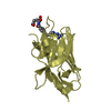
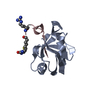
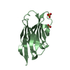
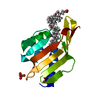
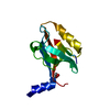
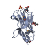
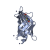
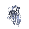
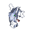
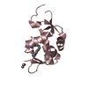
| Title | crystal structure of Escherichia coli PliG, a periplasmic lysozyme inhibitor of g-type lysozyme | ||||||
 Components Components | Inhibitor of g-type lysozyme | ||||||
 Keywords Keywords | HORMONE INHIBITOR / lysozyme inhibitor / g-type lysozyme binding | ||||||
| Function / homology |  Function and homology information Function and homology informationlysozyme inhibitor activity / defense response / outer membrane-bounded periplasmic space Similarity search - Function | ||||||
| Biological species |  | ||||||
| Method |  X-RAY DIFFRACTION / MOLECULAR REPLACEMENT / Resolution: 1.8 Å X-RAY DIFFRACTION / MOLECULAR REPLACEMENT / Resolution: 1.8 Å | ||||||
 Authors Authors | Leysen, S. / Vanheuverzwijn, S. / Van Asten, K. / Vanderkelen, L. / Michiels, C.W. / Strelkov, S.V. | ||||||
 Citation Citation | Journal: J.Struct.Biol. / Year: 2012 Title: Structural characterization of the PliG lysozyme inhibitor family. Authors: Leysen, S. / Vanderkelen, L. / Van Asten, K. / Vanheuverzwijn, S. / Theuwis, V. / Michiels, C.W. / Strelkov, S.V. | ||||||
| History |
|
- Structure visualization
| Structure viewer | Molecule: MolmilJmol/JSmol |
|---|
- Downloads & links
-Download
| PDBx/mmCIF format | 4dy3.cif.gz | 109.5 KB | Display | PDBx/mmCIF format |
|---|---|---|---|---|
| PDB format | pdb4dy3.ent.gz | 85.6 KB | Display | PDB format |
| PDBx/mmJSON format | 4dy3.json.gz | Tree view | PDBx/mmJSON format | |
| Others |  Other downloads Other downloads |
-Validation report
| Arichive directory | https://data.pdbj.org/pub/pdb/validation_reports/dy/4dy3ftp://data.pdbj.org/pub/pdb/validation_reports/dy/4dy3 | HTTPS FTP |
|---|
-Related structure data


-Links
 PDBj
PDBj- Assembly
| Deposited unit | 
| ||||||||
|---|---|---|---|---|---|---|---|---|---|
| 1 | 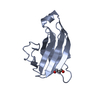
| ||||||||
| 2 | 
| ||||||||
| Unit cell |
|
-Components
| #1: Protein | Mass: 12538.815 Da / Num. of mol.: 2 / Fragment: UNP residues 23-133 Source method: isolated from a genetically manipulated source Source: (gene. exp.) #2: Chemical |   Mass: 92.094 Da / Num. of mol.: 2 / Source method: obtained synthetically / Formula: C3H8O3 Mass: 92.094 Da / Num. of mol.: 2 / Source method: obtained synthetically / Formula: C3H8O3#3: Water | ChemComp-HOH / |  Mass: 18.015 Da / Num. of mol.: 354 / Source method: isolated from a natural source / Formula: H2O Mass: 18.015 Da / Num. of mol.: 354 / Source method: isolated from a natural source / Formula: H2O |
|---|
-Experimental details
-Experiment
| Experiment | Method: X-RAY DIFFRACTION / Number of used crystals: 1 |
|---|
- Sample preparation
| Crystal | Density Matthews: 2.17 Å3/Da / Density % sol: 43.27 % |
|---|---|
| Crystal grow | Temperature: 293 K / Method: vapor diffusion, hanging drop / pH: 5 Details: 0.1M sodium acetate pH5.0, 0.15M NaCl, 25% w/v PEG6000, VAPOR DIFFUSION, HANGING DROP, temperature 293K |
-Data collection
| Diffraction | Mean temperature: 100 K |
|---|---|
| Diffraction source | Source: ROTATING ANODE / Type: RIGAKU MICROMAX-007 HF / Wavelength: 1.54 Å |
| Detector | Type: MAR scanner 345 mm plate / Detector: IMAGE PLATE / Date: Apr 8, 2010 |
| Radiation | Monochromator: mirrors / Protocol: SINGLE WAVELENGTH / Monochromatic (M) / Laue (L): M / Scattering type: x-ray |
| Radiation wavelength | Wavelength: 1.54 Å / Relative weight: 1 |
| Reflection | Resolution: 1.8→19.69 Å / Num. all: 24224 / Num. obs: 22521 / % possible obs: 93 % / Observed criterion σ(F): -3 / Observed criterion σ(I): 2 |
| Reflection shell | Resolution: 1.8→1.9 Å / Redundancy: 4.6 % / Mean I/σ(I) obs: 3.8 / Num. unique all: 2712 / Rsym value: 0.437 / % possible all: 91.1 |
- Processing
| Software |
| |||||||||||||||||||||||||||||||||||||||||||||||||||||||||||||||||||||||||||
|---|---|---|---|---|---|---|---|---|---|---|---|---|---|---|---|---|---|---|---|---|---|---|---|---|---|---|---|---|---|---|---|---|---|---|---|---|---|---|---|---|---|---|---|---|---|---|---|---|---|---|---|---|---|---|---|---|---|---|---|---|---|---|---|---|---|---|---|---|---|---|---|---|---|---|---|---|
| Refinement | Method to determine structure: MOLECULAR REPLACEMENT / Resolution: 1.8→19.69 Å / SU ML: 0.38 / σ(F): 1.34 / Phase error: 19.7 / Stereochemistry target values: ML
| |||||||||||||||||||||||||||||||||||||||||||||||||||||||||||||||||||||||||||
| Solvent computation | Shrinkage radii: 0.72 Å / VDW probe radii: 1 Å / Solvent model: FLAT BULK SOLVENT MODEL / Bsol: 47.867 Å2 / ksol: 0.413 e/Å3 | |||||||||||||||||||||||||||||||||||||||||||||||||||||||||||||||||||||||||||
| Displacement parameters |
| |||||||||||||||||||||||||||||||||||||||||||||||||||||||||||||||||||||||||||
| Refinement step | Cycle: LAST / Resolution: 1.8→19.69 Å
| |||||||||||||||||||||||||||||||||||||||||||||||||||||||||||||||||||||||||||
| Refine LS restraints |
| |||||||||||||||||||||||||||||||||||||||||||||||||||||||||||||||||||||||||||
| LS refinement shell |
| |||||||||||||||||||||||||||||||||||||||||||||||||||||||||||||||||||||||||||
| Refinement TLS params. | Method: refined / Refine-ID: X-RAY DIFFRACTION
| |||||||||||||||||||||||||||||||||||||||||||||||||||||||||||||||||||||||||||
| Refinement TLS group |
|
