 Movie
Movie Controller


+ Open data

- Basic information
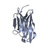
| Entry | Database: PDB / ID: 5i0z | ||||||
|---|---|---|---|---|---|---|---|
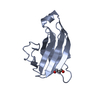
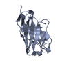
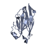
| Title | Crystal structure of the single domain catalytic antibody 3D8-VH | ||||||
 Components Components | catalytic DNA antibody | ||||||
 Keywords Keywords | IMMUNE SYSTEM / 3D8 scFv / 3D8-VH | ||||||
| Function / homology | Immunoglobulins / Immunoglobulin-like / Sandwich / Mainly Beta Function and homology information Function and homology information | ||||||
| Biological species |  | ||||||
| Method |  X-RAY DIFFRACTION / SYNCHROTRON / MOLECULAR REPLACEMENT / Resolution: 1.9 Å X-RAY DIFFRACTION / SYNCHROTRON / MOLECULAR REPLACEMENT / Resolution: 1.9 Å | ||||||
 Authors Authors | Park, S.Y. / Kim, J.S. | ||||||
| Funding support |  Korea, Republic Of, 1items Korea, Republic Of, 1items
| ||||||
 Citation Citation | Journal: To Be Published Title: Crystal structure of the single domain catalytic antibody 3D8-VH Authors: Park, S.Y. / Kim, B.N. | ||||||
| History |
|
- Structure visualization
| Structure viewer | Molecule: MolmilJmol/JSmol |
|---|
- Downloads & links
-Download
| PDBx/mmCIF format | 5i0z.cif.gz | 121.9 KB | Display | PDBx/mmCIF format |
|---|---|---|---|---|
| PDB format | pdb5i0z.ent.gz | 93.4 KB | Display | PDB format |
| PDBx/mmJSON format | 5i0z.json.gz | Tree view | PDBx/mmJSON format | |
| Others |  Other downloads Other downloads |
-Validation report
| Arichive directory | https://data.pdbj.org/pub/pdb/validation_reports/i0/5i0zftp://data.pdbj.org/pub/pdb/validation_reports/i0/5i0z | HTTPS FTP |
|---|
-Related structure data
| Related structure data |  2gkiS S: Starting model for refinement |
|---|---|
| Similar structure data |


-Links
 PDBj
PDBj

- Assembly
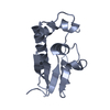
| Deposited unit | 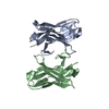
| ||||||||||||
|---|---|---|---|---|---|---|---|---|---|---|---|---|---|
| 1 | 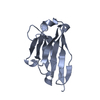
| ||||||||||||
| 2 | 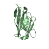
| ||||||||||||
| Unit cell |
| ||||||||||||
| Components on special symmetry positions |
|
-Components
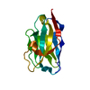
| #1: Antibody | Mass: 15814.971 Da / Num. of mol.: 2 Source method: isolated from a genetically manipulated source Source: (gene. exp.)  #2: Water | ChemComp-HOH / |  Mass: 18.015 Da / Num. of mol.: 350 / Source method: isolated from a natural source / Formula: H2O Mass: 18.015 Da / Num. of mol.: 350 / Source method: isolated from a natural source / Formula: H2OHas protein modification | Y | |
|---|
-Experimental details
-Experiment
| Experiment | Method: X-RAY DIFFRACTION / Number of used crystals: 1 |
|---|
- Sample preparation
| Crystal | Density Matthews: 1.97 Å3/Da / Density % sol: 37.42 % Description: THE ENTRY CONTAINS FRIEDEL PAIRS IN F_PLUS/MINUS COLUMNS |
|---|---|
| Crystal grow | Temperature: 295 K / Method: vapor diffusion, hanging drop / Details: 30 % (w/v) PEG 8000 and 0.1 M ammonium sulfate |
-Data collection
| Diffraction | Mean temperature: 100 K |
|---|---|
| Diffraction source | Source: SYNCHROTRON / Site: PAL/PLS / Beamline: 5C (4A) / Wavelength: 1.24 Å |
| Detector | Type: ADSC QUANTUM 315r / Detector: CCD / Date: Feb 27, 2015 |
| Radiation | Protocol: SINGLE WAVELENGTH / Monochromatic (M) / Laue (L): M / Scattering type: x-ray |
| Radiation wavelength | Wavelength: 1.24 Å / Relative weight: 1 |
| Reflection | Resolution: 1.9→50 Å / Num. obs: 16712 / % possible obs: 96.3 % / Redundancy: 2.7 % / Net I/σ(I): 6.3 |
| Reflection shell | Resolution: 1.9→2 Å |
- Processing
| Software |
| |||||||||||||||||||||||||||||||||||||||||||||||||||||||||||||||||||||||||||||||||||||||||||
|---|---|---|---|---|---|---|---|---|---|---|---|---|---|---|---|---|---|---|---|---|---|---|---|---|---|---|---|---|---|---|---|---|---|---|---|---|---|---|---|---|---|---|---|---|---|---|---|---|---|---|---|---|---|---|---|---|---|---|---|---|---|---|---|---|---|---|---|---|---|---|---|---|---|---|---|---|---|---|---|---|---|---|---|---|---|---|---|---|---|---|---|---|
| Refinement | Method to determine structure: MOLECULAR REPLACEMENT Starting model: 2GKI Resolution: 1.9→37.402 Å / SU ML: 0.24 / Cross valid method: FREE R-VALUE / σ(F): 1.35 / Phase error: 26.27 Details: THE ENTRY CONTAINS FRIEDEL PAIRS IN F_PLUS/MINUS COLUMNS
| |||||||||||||||||||||||||||||||||||||||||||||||||||||||||||||||||||||||||||||||||||||||||||
| Solvent computation | Shrinkage radii: 0.9 Å / VDW probe radii: 1.11 Å | |||||||||||||||||||||||||||||||||||||||||||||||||||||||||||||||||||||||||||||||||||||||||||
| Refinement step | Cycle: LAST / Resolution: 1.9→37.402 Å
| |||||||||||||||||||||||||||||||||||||||||||||||||||||||||||||||||||||||||||||||||||||||||||
| Refine LS restraints |
| |||||||||||||||||||||||||||||||||||||||||||||||||||||||||||||||||||||||||||||||||||||||||||
| LS refinement shell |
| |||||||||||||||||||||||||||||||||||||||||||||||||||||||||||||||||||||||||||||||||||||||||||
| Refinement TLS params. | Method: refined / Origin x: -16.2106 Å / Origin y: 3.1102 Å / Origin z: -0.0331 Å
| |||||||||||||||||||||||||||||||||||||||||||||||||||||||||||||||||||||||||||||||||||||||||||
| Refinement TLS group | Selection details: all |
