 Movie
Movie Controller

[English] 日本語
 Yorodumi
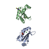
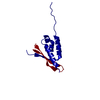
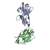
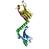
- PDB-2gki: Heavy and light chain variable single domains of an anti-DNA bind... -
+ Open data

- Basic information
| Entry | Database: PDB / ID: 2gki | ||||||
|---|---|---|---|---|---|---|---|
| Title | Heavy and light chain variable single domains of an anti-DNA binding antibody hydrolyze both double- and single-stranded DNAs without sequence specificity | ||||||
 Components Components | nuclease | ||||||
 Keywords Keywords | IMMUNE SYSTEM / anti-DNA antibody / catalytic antibody | ||||||
| Function / homology |  Function and homology information Function and homology informationimmunoglobulin complex / adaptive immune response / extracellular region Similarity search - Function | ||||||
| Biological species |  | ||||||
| Method |  X-RAY DIFFRACTION / SYNCHROTRON / MOLECULAR REPLACEMENT / Resolution: 2.88 Å X-RAY DIFFRACTION / SYNCHROTRON / MOLECULAR REPLACEMENT / Resolution: 2.88 Å | ||||||
 Authors Authors | Kim, Y.R. / Kim, J.S. / Lee, S.H. / Lee, W.R. / Sohn, J.N. / Chung, Y.C. / Shim, H.K. / Lee, S.C. / Kwon, M.H. / Kim, Y.S. | ||||||
 Citation Citation | Journal: J.Biol.Chem. / Year: 2006 Title: Heavy and light chain variable single domains of an anti-DNA binding antibody hydrolyze both double- and single-stranded DNAs without sequence specificity. Authors: Kim, Y.R. / Kim, J.S. / Lee, S.H. / Lee, W.R. / Sohn, J.N. / Chung, Y.C. / Shim, H.K. / Lee, S.C. / Kwon, M.H. / Kim, Y.S. | ||||||
| History |
|
- Structure visualization
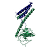
| Structure viewer | Molecule: MolmilJmol/JSmol |
|---|
- Downloads & links
-Download
| PDBx/mmCIF format | 2gki.cif.gz | 106.4 KB | Display | PDBx/mmCIF format |
|---|---|---|---|---|
| PDB format | pdb2gki.ent.gz | 82.9 KB | Display | PDB format |
| PDBx/mmJSON format | 2gki.json.gz | Tree view | PDBx/mmJSON format | |
| Others |  Other downloads Other downloads |
-Validation report
| Arichive directory | https://data.pdbj.org/pub/pdb/validation_reports/gk/2gkiftp://data.pdbj.org/pub/pdb/validation_reports/gk/2gki | HTTPS FTP |
|---|
-Related structure data
| Related structure data | 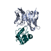 1dzbS S: Starting model for refinement |
|---|---|
| Similar structure data |

-Links
 PDBj
PDBj

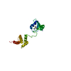
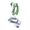
- Assembly
| Deposited unit | 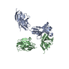
| ||||||||
|---|---|---|---|---|---|---|---|---|---|
| 1 | 
| ||||||||
| 2 | 
| ||||||||
| 3 | 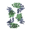
| ||||||||
| 4 | 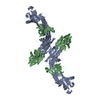
| ||||||||
| Unit cell |
|
-Components
| #1: Antibody | Mass: 31504.314 Da / Num. of mol.: 2 Source method: isolated from a genetically manipulated source Source: (gene. exp.)  References: GenBank: 8777863, GenBank: 8777865, UniProt: A0N274*PLUS #2: Water | ChemComp-HOH / |  Mass: 18.015 Da / Num. of mol.: 70 / Source method: isolated from a natural source / Formula: H2O Mass: 18.015 Da / Num. of mol.: 70 / Source method: isolated from a natural source / Formula: H2OHas protein modification | Y | |
|---|
-Experimental details
-Experiment
| Experiment | Method: X-RAY DIFFRACTION / Number of used crystals: 1 |
|---|
- Sample preparation
| Crystal | Density Matthews: 6.814426 Å3/Da / Density % sol: 81.950058 % |
|---|---|
| Crystal grow | Temperature: 291 K / Method: vapor diffusion, hanging drop / pH: 7 Details: 2M Lithium sulfate, pH 7.0, VAPOR DIFFUSION, HANGING DROP, temperature 291.0K |
-Data collection
| Diffraction | Mean temperature: 100 K |
|---|---|
| Diffraction source | Source: SYNCHROTRON / Site: PAL/PLS  / Beamline: 4A / Wavelength: 1 Å / Beamline: 4A / Wavelength: 1 Å |
| Detector | Type: ADSC QUANTUM 315 / Detector: CCD / Date: May 22, 2005 |
| Radiation | Monochromator: GRAPHITE / Protocol: SINGLE WAVELENGTH / Monochromatic (M) / Laue (L): M / Scattering type: x-ray |
| Radiation wavelength | Wavelength: 1 Å / Relative weight: 1 |
| Reflection | Resolution: 2.88→100 Å / Num. all: 40011 / Num. obs: 40011 / % possible obs: 99.4 % / Observed criterion σ(F): -3 / Observed criterion σ(I): -3 / Redundancy: 11.8 % / Rmerge(I) obs: 0.078 |
| Reflection shell | Resolution: 2.88→2.98 Å / % possible all: 99.6 |
- Processing
| Software |
| |||||||||||||||||||||||||
|---|---|---|---|---|---|---|---|---|---|---|---|---|---|---|---|---|---|---|---|---|---|---|---|---|---|---|
| Refinement | Method to determine structure: MOLECULAR REPLACEMENT Starting model: 1DZB Resolution: 2.88→49.58 Å / σ(F): -3 / Stereochemistry target values: Engh & Huber
| |||||||||||||||||||||||||
| Refinement step | Cycle: LAST / Resolution: 2.88→49.58 Å
|
