 Movie
Movie Controller

[English] 日本語
 Yorodumi
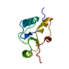
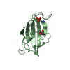
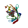
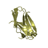
- PDB-5wbh: Structure of the FRB domain of mTOR bound to a substrate recruitm... -
+ Open data

- Basic information
| Entry | Database: PDB / ID: 5wbh | ||||||
|---|---|---|---|---|---|---|---|
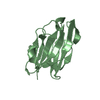
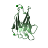
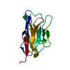
| Title | Structure of the FRB domain of mTOR bound to a substrate recruitment peptide of S6K1 | ||||||
 Components Components |
| ||||||
 Keywords Keywords | TRANSFERASE | ||||||
| Function / homology |  Function and homology information Function and homology informationlong-chain fatty acid import into cell / positive regulation of SCF-dependent proteasomal ubiquitin-dependent catabolic process / RNA polymerase III type 2 promoter sequence-specific DNA binding / RNA polymerase III type 1 promoter sequence-specific DNA binding / positive regulation of cytoplasmic translational initiation / regulation of locomotor rhythm / T-helper 1 cell lineage commitment / positive regulation of pentose-phosphate shunt / positive regulation of wound healing, spreading of epidermal cells / regulation of membrane permeability ...long-chain fatty acid import into cell / positive regulation of SCF-dependent proteasomal ubiquitin-dependent catabolic process / RNA polymerase III type 2 promoter sequence-specific DNA binding / RNA polymerase III type 1 promoter sequence-specific DNA binding / positive regulation of cytoplasmic translational initiation / regulation of locomotor rhythm / T-helper 1 cell lineage commitment / positive regulation of pentose-phosphate shunt / positive regulation of wound healing, spreading of epidermal cells / regulation of membrane permeability / TORC2 complex / cellular response to leucine starvation / TFIIIC-class transcription factor complex binding / heart valve morphogenesis / voluntary musculoskeletal movement / negative regulation of TORC2 signaling / negative regulation of lysosome organization / TORC1 complex / calcineurin-NFAT signaling cascade / RNA polymerase III type 3 promoter sequence-specific DNA binding / positive regulation of transcription of nucleolar large rRNA by RNA polymerase I / positive regulation of keratinocyte migration / regulation of osteoclast differentiation / energy reserve metabolic process / MTOR signalling / regulation of lysosome organization / cellular response to L-leucine / Energy dependent regulation of mTOR by LKB1-AMPK / cellular response to nutrient / regulation of autophagosome assembly / Amino acids regulate mTORC1 / cellular response to methionine / negative regulation of cell size / TORC2 signaling / cellular response to osmotic stress / cell projection organization / anoikis / inositol hexakisphosphate binding / cardiac muscle cell development / negative regulation of calcineurin-NFAT signaling cascade / positive regulation of ubiquitin-dependent protein catabolic process / regulation of myelination / negative regulation of protein localization to nucleus / positive regulation of transcription by RNA polymerase III / positive regulation of ruffle assembly / regulation of cell size / positive regulation of myotube differentiation / negative regulation of macroautophagy / Macroautophagy / Constitutive Signaling by AKT1 E17K in Cancer / phosphatidylinositol-mediated signaling / positive regulation of actin filament polymerization / germ cell development / oligodendrocyte differentiation / TORC1 signaling / positive regulation of oligodendrocyte differentiation / behavioral response to pain / response to amino acid / TOR signaling / mTORC1-mediated signalling / CD28 dependent PI3K/Akt signaling / HSF1-dependent transactivation / regulation of macroautophagy / cellular response to dexamethasone stimulus / positive regulation of translational initiation / positive regulation of epithelial to mesenchymal transition / positive regulation of lipid biosynthetic process / 'de novo' pyrimidine nucleobase biosynthetic process / behavioral fear response / vascular endothelial cell response to laminar fluid shear stress / heart morphogenesis / regulation of cellular response to heat / positive regulation of lamellipodium assembly / neuronal action potential / T cell costimulation / phagocytic vesicle / positive regulation of stress fiber assembly / cardiac muscle contraction / cytoskeleton organization / protein serine/threonine/tyrosine kinase activity / negative regulation of insulin receptor signaling pathway / positive regulation of TORC1 signaling / endomembrane system / positive regulation of mitotic cell cycle / cellular response to nutrient levels / positive regulation of glycolytic process / cellular response to amino acid starvation / cellular response to starvation / peptidyl-serine phosphorylation / regulation of signal transduction by p53 class mediator / Regulation of PTEN gene transcription / negative regulation of autophagy / negative regulation of extrinsic apoptotic signaling pathway / VEGFR2 mediated vascular permeability / post-embryonic development / TP53 Regulates Metabolic Genes / regulation of actin cytoskeleton organization / non-specific protein-tyrosine kinase / positive regulation of translation / macroautophagy Similarity search - Function | ||||||
| Biological species |  Homo sapiens (human) Homo sapiens (human) | ||||||
| Method |  X-RAY DIFFRACTION / SYNCHROTRON / MOLECULAR REPLACEMENT / Resolution: 1.75 Å X-RAY DIFFRACTION / SYNCHROTRON / MOLECULAR REPLACEMENT / Resolution: 1.75 Å | ||||||
 Authors Authors | Pavletich, N.P. / Yang, H. | ||||||
 Citation Citation | Journal: Nature / Year: 2017 Title: Mechanisms of mTORC1 activation by RHEB and inhibition by PRAS40. Authors: Haijuan Yang / Xiaolu Jiang / Buren Li / Hyo J Yang / Meredith Miller / Angela Yang / Ankita Dhar / Nikola P Pavletich /  Abstract: The mechanistic target of rapamycin complex 1 (mTORC1) controls cell growth and metabolism in response to nutrients, energy levels, and growth factors. It contains the atypical kinase mTOR and the ...The mechanistic target of rapamycin complex 1 (mTORC1) controls cell growth and metabolism in response to nutrients, energy levels, and growth factors. It contains the atypical kinase mTOR and the RAPTOR subunit that binds to the Tor signalling sequence (TOS) motif of substrates and regulators. mTORC1 is activated by the small GTPase RHEB (Ras homologue enriched in brain) and inhibited by PRAS40. Here we present the 3.0 ångström cryo-electron microscopy structure of mTORC1 and the 3.4 ångström structure of activated RHEB-mTORC1. RHEB binds to mTOR distally from the kinase active site, yet causes a global conformational change that allosterically realigns active-site residues, accelerating catalysis. Cancer-associated hyperactivating mutations map to structural elements that maintain the inactive state, and we provide biochemical evidence that they mimic RHEB relieving auto-inhibition. We also present crystal structures of RAPTOR-TOS motif complexes that define the determinants of TOS recognition, of an mTOR FKBP12-rapamycin-binding (FRB) domain-substrate complex that establishes a second substrate-recruitment mechanism, and of a truncated mTOR-PRAS40 complex that reveals PRAS40 inhibits both substrate-recruitment sites. These findings help explain how mTORC1 selects its substrates, how its kinase activity is controlled, and how it is activated by cancer-associated mutations. | ||||||
| History |
|
- Structure visualization
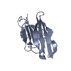
| Structure viewer | Molecule: MolmilJmol/JSmol |
|---|
- Downloads & links
-Download
| PDBx/mmCIF format | 5wbh.cif.gz | 404.1 KB | Display | PDBx/mmCIF format |
|---|---|---|---|---|
| PDB format | pdb5wbh.ent.gz | 333.8 KB | Display | PDB format |
| PDBx/mmJSON format | 5wbh.json.gz | Tree view | PDBx/mmJSON format | |
| Others |  Other downloads Other downloads |
-Validation report
| Arichive directory | https://data.pdbj.org/pub/pdb/validation_reports/wb/5wbhftp://data.pdbj.org/pub/pdb/validation_reports/wb/5wbh | HTTPS FTP |
|---|
-Related structure data
| Related structure data |  7086C  7087C  5wbiC  5wbjC  5wbkC  5wblC  5wbuC  5wbyC  6bcuC 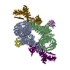 6bcxC  1fapS S: Starting model for refinement C: citing same article ( |
|---|---|
| Similar structure data |

-Links
 PDBj
PDBj


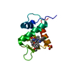
- Assembly
| Deposited unit | 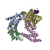
| ||||||||
|---|---|---|---|---|---|---|---|---|---|
| 1 | 
| ||||||||
| 2 | 
| ||||||||
| 3 | 
| ||||||||
| 4 | 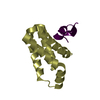
| ||||||||
| 5 | 
| ||||||||
| Unit cell |
|
-Components
| #1: Protein | Mass: 12091.749 Da / Num. of mol.: 5 / Fragment: residues 2018-2114 Source method: isolated from a genetically manipulated source Source: (gene. exp.) Homo sapiens (human) / Gene: MTOR, FRAP, FRAP1, FRAP2, RAFT1, RAPT1 / Plasmid: pET26 / Production host:  References: UniProt: P42345, non-specific serine/threonine protein kinase #2: Protein/peptide | | Mass: 3057.544 Da / Num. of mol.: 1 / Fragment: residues 412-437 Source method: isolated from a genetically manipulated source Source: (gene. exp.) Homo sapiens (human) / Gene: RPS6KB1, STK14A / Plasmid: pET26 / Production host: References: UniProt: P23443, non-specific serine/threonine protein kinase #3: Water | ChemComp-HOH / |  Mass: 18.015 Da / Num. of mol.: 334 / Source method: isolated from a natural source / Formula: H2O Mass: 18.015 Da / Num. of mol.: 334 / Source method: isolated from a natural source / Formula: H2O |
|---|
-Experimental details
-Experiment
| Experiment | Method: X-RAY DIFFRACTION / Number of used crystals: 1 |
|---|
- Sample preparation
| Crystal | Density Matthews: 2.77 Å3/Da / Density % sol: 55.58 % / Mosaicity: 0.738 ° |
|---|---|
| Crystal grow | Temperature: 289 K / Method: vapor diffusion, hanging drop / pH: 7 / Details: tacsimate |
-Data collection
| Diffraction | Mean temperature: 100 K | ||||||||||||||||||||||||||||||||||||||||||||||||||||||||||||||||||||||||||||||||||||||||||||||||||||||||||||||
|---|---|---|---|---|---|---|---|---|---|---|---|---|---|---|---|---|---|---|---|---|---|---|---|---|---|---|---|---|---|---|---|---|---|---|---|---|---|---|---|---|---|---|---|---|---|---|---|---|---|---|---|---|---|---|---|---|---|---|---|---|---|---|---|---|---|---|---|---|---|---|---|---|---|---|---|---|---|---|---|---|---|---|---|---|---|---|---|---|---|---|---|---|---|---|---|---|---|---|---|---|---|---|---|---|---|---|---|---|---|---|---|
| Diffraction source | Source: SYNCHROTRON / Site: APS / Beamline: 24-ID-C / Wavelength: 0.9792 Å | ||||||||||||||||||||||||||||||||||||||||||||||||||||||||||||||||||||||||||||||||||||||||||||||||||||||||||||||
| Detector | Type: ADSC QUANTUM 315 / Detector: CCD / Date: Feb 12, 2014 | ||||||||||||||||||||||||||||||||||||||||||||||||||||||||||||||||||||||||||||||||||||||||||||||||||||||||||||||
| Radiation | Protocol: SINGLE WAVELENGTH / Monochromatic (M) / Laue (L): M / Scattering type: x-ray | ||||||||||||||||||||||||||||||||||||||||||||||||||||||||||||||||||||||||||||||||||||||||||||||||||||||||||||||
| Radiation wavelength | Wavelength: 0.9792 Å / Relative weight: 1 | ||||||||||||||||||||||||||||||||||||||||||||||||||||||||||||||||||||||||||||||||||||||||||||||||||||||||||||||
| Reflection | Resolution: 1.75→50 Å / Num. obs: 66487 / % possible obs: 98.2 % / Redundancy: 4.9 % / Rmerge(I) obs: 0.068 / Rpim(I) all: 0.032 / Rrim(I) all: 0.076 / Χ2: 1.575 / Net I/σ(I): 11.6 / Num. measured all: 328879 | ||||||||||||||||||||||||||||||||||||||||||||||||||||||||||||||||||||||||||||||||||||||||||||||||||||||||||||||
| Reflection shell | Diffraction-ID: 1
|
- Processing
| Software |
| |||||||||||||||||||||||||||||||||||||||||||||||||||||||||||||||||||||||||||||||||||||||||||||||||||||||||||||||||||||||||||||||||||||||||||||||||||||||||||||||||||||||||||||||
|---|---|---|---|---|---|---|---|---|---|---|---|---|---|---|---|---|---|---|---|---|---|---|---|---|---|---|---|---|---|---|---|---|---|---|---|---|---|---|---|---|---|---|---|---|---|---|---|---|---|---|---|---|---|---|---|---|---|---|---|---|---|---|---|---|---|---|---|---|---|---|---|---|---|---|---|---|---|---|---|---|---|---|---|---|---|---|---|---|---|---|---|---|---|---|---|---|---|---|---|---|---|---|---|---|---|---|---|---|---|---|---|---|---|---|---|---|---|---|---|---|---|---|---|---|---|---|---|---|---|---|---|---|---|---|---|---|---|---|---|---|---|---|---|---|---|---|---|---|---|---|---|---|---|---|---|---|---|---|---|---|---|---|---|---|---|---|---|---|---|---|---|---|---|---|---|---|
| Refinement | Method to determine structure: MOLECULAR REPLACEMENT Starting model: 1FAP Resolution: 1.75→50.01 Å / Cor.coef. Fo:Fc: 0.962 / Cor.coef. Fo:Fc free: 0.955 / SU B: 7.489 / SU ML: 0.102 / Cross valid method: THROUGHOUT / σ(F): 0 / ESU R: 0.111 / ESU R Free: 0.102 Details: U VALUES : WITH TLS ADDED HYDROGENS HAVE BEEN USED IF PRESENT IN THE INPUT
| |||||||||||||||||||||||||||||||||||||||||||||||||||||||||||||||||||||||||||||||||||||||||||||||||||||||||||||||||||||||||||||||||||||||||||||||||||||||||||||||||||||||||||||||
| Solvent computation | Ion probe radii: 0.8 Å / Shrinkage radii: 0.8 Å / VDW probe radii: 1.2 Å | |||||||||||||||||||||||||||||||||||||||||||||||||||||||||||||||||||||||||||||||||||||||||||||||||||||||||||||||||||||||||||||||||||||||||||||||||||||||||||||||||||||||||||||||
| Displacement parameters | Biso max: 130.28 Å2 / Biso mean: 37.909 Å2 / Biso min: 12.88 Å2
| |||||||||||||||||||||||||||||||||||||||||||||||||||||||||||||||||||||||||||||||||||||||||||||||||||||||||||||||||||||||||||||||||||||||||||||||||||||||||||||||||||||||||||||||
| Refinement step | Cycle: final / Resolution: 1.75→50.01 Å
| |||||||||||||||||||||||||||||||||||||||||||||||||||||||||||||||||||||||||||||||||||||||||||||||||||||||||||||||||||||||||||||||||||||||||||||||||||||||||||||||||||||||||||||||
| Refine LS restraints |
| |||||||||||||||||||||||||||||||||||||||||||||||||||||||||||||||||||||||||||||||||||||||||||||||||||||||||||||||||||||||||||||||||||||||||||||||||||||||||||||||||||||||||||||||
| LS refinement shell | Resolution: 1.75→1.795 Å / Rfactor Rfree error: 0 / Total num. of bins used: 20
| |||||||||||||||||||||||||||||||||||||||||||||||||||||||||||||||||||||||||||||||||||||||||||||||||||||||||||||||||||||||||||||||||||||||||||||||||||||||||||||||||||||||||||||||
| Refinement TLS params. | Method: refined / Refine-ID: X-RAY DIFFRACTION
| |||||||||||||||||||||||||||||||||||||||||||||||||||||||||||||||||||||||||||||||||||||||||||||||||||||||||||||||||||||||||||||||||||||||||||||||||||||||||||||||||||||||||||||||
| Refinement TLS group |
|
