 Movie
Movie Controller


+ Open data

- Basic information
| Entry | Database: PDB / ID: 3ft1 | ||||||
|---|---|---|---|---|---|---|---|
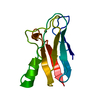
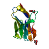
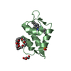
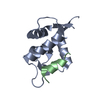
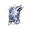
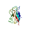
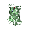
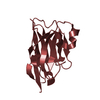
| Title | Crystal structure of pollen allergen Phl p 3 | ||||||
 Components Components | Phl p 3 allergen | ||||||
 Keywords Keywords | ALLERGEN / BETA-BARREL | ||||||
| Function / homology |  Function and homology information Function and homology information | ||||||
| Biological species |  Phleum pratense (timothy grass) Phleum pratense (timothy grass) | ||||||
| Method |  X-RAY DIFFRACTION / SYNCHROTRON / MOLECULAR REPLACEMENT / Resolution: 1.79 Å X-RAY DIFFRACTION / SYNCHROTRON / MOLECULAR REPLACEMENT / Resolution: 1.79 Å | ||||||
 Authors Authors | Keller, W. / Devanaboyina, S.C. | ||||||
 Citation Citation | Journal: Allergy / Year: 2014 Title: High-resolution crystal structure and IgE recognition of the major grass pollen allergen Phl p 3. Authors: Devanaboyina, S.C. / Cornelius, C. / Lupinek, C. / Fauland, K. / Dall'Antonia, F. / Nandy, A. / Hagen, S. / Flicker, S. / Valenta, R. / Keller, W. | ||||||
| History |
|
- Structure visualization
| Structure viewer | Molecule: MolmilJmol/JSmol |
|---|
- Downloads & links
-Download
| PDBx/mmCIF format | 3ft1.cif.gz | 104.1 KB | Display | PDBx/mmCIF format |
|---|---|---|---|---|
| PDB format | pdb3ft1.ent.gz | 79.3 KB | Display | PDB format |
| PDBx/mmJSON format | 3ft1.json.gz | Tree view | PDBx/mmJSON format | |
| Others |  Other downloads Other downloads |
-Validation report
| Arichive directory | https://data.pdbj.org/pub/pdb/validation_reports/ft/3ft1ftp://data.pdbj.org/pub/pdb/validation_reports/ft/3ft1 | HTTPS FTP |
|---|
-Related structure data
| Related structure data |  3ft9C  1whoS S: Starting model for refinement C: citing same article ( |
|---|---|
| Similar structure data |


-Links
 PDBj
PDBj- Assembly
| Deposited unit | 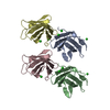
| ||||||||
|---|---|---|---|---|---|---|---|---|---|
| 1 | 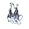
| ||||||||
| 2 | 
| ||||||||
| 3 | 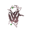
| ||||||||
| 4 | 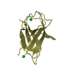
| ||||||||
| Unit cell |
|
-Components
| #1: Protein | Mass: 11378.952 Da / Num. of mol.: 4 Source method: isolated from a genetically manipulated source Source: (gene. exp.) Phleum pratense (timothy grass) / Production host:  #2: Chemical | ChemComp-CL /   Mass: 35.453 Da / Num. of mol.: 10 / Source method: obtained synthetically / Formula: Cl Mass: 35.453 Da / Num. of mol.: 10 / Source method: obtained synthetically / Formula: Cl#3: Water | ChemComp-HOH / |  Mass: 18.015 Da / Num. of mol.: 571 / Source method: isolated from a natural source / Formula: H2O Mass: 18.015 Da / Num. of mol.: 571 / Source method: isolated from a natural source / Formula: H2O |
|---|
-Experimental details
-Experiment
| Experiment | Method: X-RAY DIFFRACTION / Number of used crystals: 1 |
|---|
- Sample preparation
| Crystal | Density Matthews: 2.07 Å3/Da / Density % sol: 40.49 % |
|---|---|
| Crystal grow | Temperature: 293 K / Method: vapor diffusion, sitting drop / pH: 5.5 Details: 25% PEG 3350, 0.1 M Bis-tris, 0.1 M NaCl, pH 5.5, VAPOR DIFFUSION, SITTING DROP, temperature 293K |
-Data collection
| Diffraction | Mean temperature: 100 K |
|---|---|
| Diffraction source | Source: SYNCHROTRON / Site: EMBL/DESY, HAMBURG  / Beamline: X12 / Wavelength: 0.8148 Å / Beamline: X12 / Wavelength: 0.8148 Å |
| Detector | Type: MARMOSAIC 225 mm CCD / Detector: CCD / Date: Jul 2, 2007 |
| Radiation | Monochromator: Double crystal Si[111], horizontally focussing Protocol: SINGLE WAVELENGTH / Monochromatic (M) / Laue (L): M / Scattering type: x-ray |
| Radiation wavelength | Wavelength: 0.8148 Å / Relative weight: 1 |
| Reflection | Resolution: 1.79→30 Å / Num. all: 34582 / Num. obs: 34582 / % possible obs: 99.8 % / Observed criterion σ(F): 2 / Observed criterion σ(I): 2 / Redundancy: 4 % / Biso Wilson estimate: 18.3 Å2 / Rmerge(I) obs: 0.073 / Rsym value: 0.073 / Net I/σ(I): 149.7 |
| Reflection shell | Highest resolution: 1.79 Å |
- Processing
| Software |
| |||||||||||||||||||||||||
|---|---|---|---|---|---|---|---|---|---|---|---|---|---|---|---|---|---|---|---|---|---|---|---|---|---|---|
| Refinement | Method to determine structure: MOLECULAR REPLACEMENT Starting model: PDB ENTRY 1WHO Resolution: 1.79→30 Å / σ(F): 2 / σ(I): 2
| |||||||||||||||||||||||||
| Displacement parameters | Biso mean: 18.3 Å2 | |||||||||||||||||||||||||
| Refinement step | Cycle: LAST / Resolution: 1.79→30 Å
| |||||||||||||||||||||||||
| Refine LS restraints |
| |||||||||||||||||||||||||
| LS refinement shell | Highest resolution: 1.79 Å / Num. reflection obs: 206729 |
