 Movie
Movie Controller


+ Open data

- Basic information
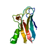
| Entry | Database: PDB / ID: 1f2x | ||||||
|---|---|---|---|---|---|---|---|
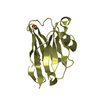
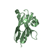
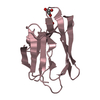
| Title | STRUCTURE OF THE SINGLE-DOMAIN CAMELID ANTIBODY CAB-CA05 | ||||||
 Components Components | ANTIBODY HEAVY CHAIN | ||||||
 Keywords Keywords | IMMUNE SYSTEM / immunoglobulin fold | ||||||
| Function / homology | Immunoglobulins / Immunoglobulin-like / Sandwich / Mainly Beta Function and homology information Function and homology information | ||||||
| Biological species |  | ||||||
| Method |  X-RAY DIFFRACTION / MOLECULAR REPLACEMENT / Resolution: 2.1 Å X-RAY DIFFRACTION / MOLECULAR REPLACEMENT / Resolution: 2.1 Å | ||||||
 Authors Authors | Decanniere, K. / Muyldermans, S. / Wyns, L. | ||||||
 Citation Citation | Journal: J.Mol.Biol. / Year: 2000 Title: Canonical antigen-binding loop structures in immunoglobulins: more structures, more canonical classes? Authors: Decanniere, K. / Muyldermans, S. / Wyns, L. #1: Journal: To be PublishedTitle: Structure of the Camelid Single-Domain Antibody Fragment cAb-CA05: Free and Complexed Structure Authors: Desmeyter, A. / Decanniere, K. / Muyldermans, S. / Wyns, L. | ||||||
| History |
|
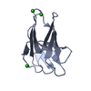
- Structure visualization
| Structure viewer | Molecule: MolmilJmol/JSmol |
|---|
- Downloads & links
-Download
| PDBx/mmCIF format | 1f2x.cif.gz | 62.1 KB | Display | PDBx/mmCIF format |
|---|---|---|---|---|
| PDB format | pdb1f2x.ent.gz | 44.9 KB | Display | PDB format |
| PDBx/mmJSON format | 1f2x.json.gz | Tree view | PDBx/mmJSON format | |
| Others |  Other downloads Other downloads |
-Validation report
| Arichive directory | https://data.pdbj.org/pub/pdb/validation_reports/f2/1f2xftp://data.pdbj.org/pub/pdb/validation_reports/f2/1f2x | HTTPS FTP |
|---|
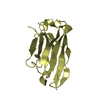
-Related structure data
| Related structure data | 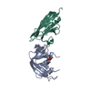 1bzqS S: Starting model for refinement |
|---|---|
| Similar structure data |


-Links
 PDBj
PDBj

- Assembly
| Deposited unit | 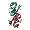
| ||||||||
|---|---|---|---|---|---|---|---|---|---|
| 1 | 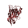
| ||||||||
| 2 | 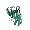
| ||||||||
| Unit cell |
|
-Components
| #1: Antibody | Mass: 14733.291 Da / Num. of mol.: 2 Source method: isolated from a genetically manipulated source Source: (gene. exp.)  #2: Water | ChemComp-HOH / |  Mass: 18.015 Da / Num. of mol.: 117 / Source method: isolated from a natural source / Formula: H2O Mass: 18.015 Da / Num. of mol.: 117 / Source method: isolated from a natural source / Formula: H2OHas protein modification | Y | |
|---|
-Experimental details
-Experiment
| Experiment | Method: X-RAY DIFFRACTION / Number of used crystals: 1 |
|---|
- Sample preparation
| Crystal | Density Matthews: 1.96 Å3/Da / Density % sol: 37.21 % | ||||||||||||||||||||
|---|---|---|---|---|---|---|---|---|---|---|---|---|---|---|---|---|---|---|---|---|---|
| Crystal grow | Temperature: 293 K / Method: vapor diffusion, hanging drop / pH: 5.6 Details: 25 % (w/v) PEG8000, 0.1 M Na citrate pH 5.6, protein concentration 1.7 mg/ml, VAPOR DIFFUSION, HANGING DROP, temperature 293K | ||||||||||||||||||||
| Crystal | *PLUS Density % sol: 45 % | ||||||||||||||||||||
| Crystal grow | *PLUS pH: 6.4 / Details: Desmyter, A., (1996) Nature Struct. Biol., 3, 803. | ||||||||||||||||||||
| Components of the solutions | *PLUS
|
-Data collection
| Diffraction | Mean temperature: 293 K |
|---|---|
| Diffraction source | Source: ROTATING ANODE / Type: RIGAKU RU200 / Wavelength: 1.5418 |
| Detector | Type: MARRESEARCH / Detector: IMAGE PLATE / Date: Jan 5, 1999 |
| Radiation | Protocol: SINGLE WAVELENGTH / Monochromatic (M) / Laue (L): M / Scattering type: x-ray |
| Radiation wavelength | Wavelength: 1.5418 Å / Relative weight: 1 |
| Reflection | Resolution: 2.1→31 Å / Num. all: 13523 / Num. obs: 13256 / % possible obs: 99.8 % / Observed criterion σ(F): 0 / Observed criterion σ(I): 0 / Redundancy: 5.8 % / Biso Wilson estimate: 23.6 Å2 / Rmerge(I) obs: 0.103 / Net I/σ(I): 4.9 |
| Reflection shell | Resolution: 2.1→2.21 Å / Redundancy: 5.3 % / Rmerge(I) obs: 0.227 / Mean I/σ(I) obs: 2.7 / Num. unique all: 1878 / % possible all: 99.8 |
- Processing
| Software |
| ||||||||||||||||||||||||||||||||||||||||||||||||
|---|---|---|---|---|---|---|---|---|---|---|---|---|---|---|---|---|---|---|---|---|---|---|---|---|---|---|---|---|---|---|---|---|---|---|---|---|---|---|---|---|---|---|---|---|---|---|---|---|---|
| Refinement | Method to determine structure: MOLECULAR REPLACEMENT Starting model: 1BZQ Resolution: 2.1→31 Å / SU B: 5.7 / SU ML: 0.15 / σ(F): 0 / σ(I): 0 / ESU R: 0.27 / ESU R Free: 0.23 / Stereochemistry target values: Engh & Huber / Details: maximum likelihood target using F's
| ||||||||||||||||||||||||||||||||||||||||||||||||
| Refinement step | Cycle: LAST / Resolution: 2.1→31 Å
| ||||||||||||||||||||||||||||||||||||||||||||||||
| Refine LS restraints |
| ||||||||||||||||||||||||||||||||||||||||||||||||
| Software | *PLUS Name: REFMAC / Classification: refinement | ||||||||||||||||||||||||||||||||||||||||||||||||
| Refinement | *PLUS Highest resolution: 2.1 Å / σ(F): 0 / % reflection Rfree: 5 % / Rfactor obs: 0.19 / Rfactor Rfree: 0.27 / Rfactor Rwork: 0.19 | ||||||||||||||||||||||||||||||||||||||||||||||||
| Solvent computation | *PLUS | ||||||||||||||||||||||||||||||||||||||||||||||||
| Displacement parameters | *PLUS | ||||||||||||||||||||||||||||||||||||||||||||||||
| Refine LS restraints | *PLUS
|
