 Movie
Movie Controller


+ Open data

- Basic information
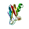
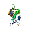
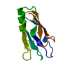
| Entry | Database: PDB / ID: 1kdj | ||||||
|---|---|---|---|---|---|---|---|
| Title | OXIDIZED FORM OF PLASTOCYANIN FROM DRYOPTERIS CRASSIRHIZOMA | ||||||
 Components Components | PLASTOCYANIN | ||||||
 Keywords Keywords | ELECTRON TRANSFER / PHOTOSYSTEM / PAI-PAI STACKING | ||||||
| Function / homology |  Function and homology information Function and homology informationchloroplast thylakoid membrane / electron transfer activity / copper ion binding Similarity search - Function | ||||||
| Biological species |  Adiantum capillus-veneris (maidenhair fern) Adiantum capillus-veneris (maidenhair fern) | ||||||
| Method |  X-RAY DIFFRACTION / SYNCHROTRON / MOLECULAR REPLACEMENT / Resolution: 1.7 Å X-RAY DIFFRACTION / SYNCHROTRON / MOLECULAR REPLACEMENT / Resolution: 1.7 Å | ||||||
 Authors Authors | Inoue, T. / Gotowda, M. / Hamada, K. / Kohzuma, T. / Yoshizaki, F. / Sugimura, Y. / Kai, Y. | ||||||
 Citation Citation | Journal: J.Biol.Chem. / Year: 1999 Title: The structure and unusual pH dependence of plastocyanin from the fern Dryopteris crassirhizoma. The protonation of an active site histidine is hindered by pi-pi interactions. Authors: Kohzuma, T. / Inoue, T. / Yoshizaki, F. / Sasakawa, Y. / Onodera, K. / Nagatomo, S. / Kitagawa, T. / Uzawa, S. / Isobe, Y. / Sugimura, Y. / Gotowda, M. / Kai, Y. | ||||||
| History |
|
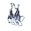
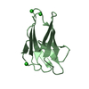
- Structure visualization
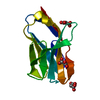
| Structure viewer | Molecule: MolmilJmol/JSmol |
|---|
- Downloads & links
-Download
| PDBx/mmCIF format | 1kdj.cif.gz | 31.3 KB | Display | PDBx/mmCIF format |
|---|---|---|---|---|
| PDB format | pdb1kdj.ent.gz | 20.9 KB | Display | PDB format |
| PDBx/mmJSON format | 1kdj.json.gz | Tree view | PDBx/mmJSON format | |
| Others |  Other downloads Other downloads |
-Validation report
| Arichive directory | https://data.pdbj.org/pub/pdb/validation_reports/kd/1kdjftp://data.pdbj.org/pub/pdb/validation_reports/kd/1kdj | HTTPS FTP |
|---|
-Related structure data


-Links
 PDBj
PDBj- Assembly
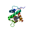
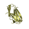
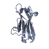
| Deposited unit | 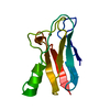
| ||||||||
|---|---|---|---|---|---|---|---|---|---|
| 1 |
| ||||||||
| Unit cell |
|
-Components
| #1: Protein | Mass: 10785.973 Da / Num. of mol.: 1 / Source method: isolated from a natural source / Details: OXIDIZED Source: (natural) Adiantum capillus-veneris (maidenhair fern)References: UniProt: Q7SIB8 |
|---|---|
| #2: Chemical | ChemComp-CU /   Mass: 63.546 Da / Num. of mol.: 1 / Source method: obtained synthetically / Formula: Cu Mass: 63.546 Da / Num. of mol.: 1 / Source method: obtained synthetically / Formula: Cu |
| #3: Water | ChemComp-HOH /  Mass: 18.015 Da / Num. of mol.: 49 / Source method: isolated from a natural source / Formula: H2O Mass: 18.015 Da / Num. of mol.: 49 / Source method: isolated from a natural source / Formula: H2O |
-Experimental details
-Experiment
| Experiment | Method: X-RAY DIFFRACTION / Number of used crystals: 1 |
|---|
- Sample preparation
| Crystal | Density Matthews: 2.4 Å3/Da / Density % sol: 48 % | ||||||||||||||||||||||||||||||
|---|---|---|---|---|---|---|---|---|---|---|---|---|---|---|---|---|---|---|---|---|---|---|---|---|---|---|---|---|---|---|---|
| Crystal grow | pH: 4.5 / Details: pH 4.5 | ||||||||||||||||||||||||||||||
| Crystal | *PLUS Density % sol: 49 % | ||||||||||||||||||||||||||||||
| Crystal grow | *PLUS Temperature: 20 ℃ / Method: vapor diffusion, hanging drop | ||||||||||||||||||||||||||||||
| Components of the solutions | *PLUS
|
-Data collection
| Diffraction | Mean temperature: 298 K |
|---|---|
| Diffraction source | Source: SYNCHROTRON / Site: Photon Factory  / Beamline: BL-6B / Wavelength: 1 / Beamline: BL-6B / Wavelength: 1 |
| Detector | Type: WEISSENBERG / Detector: DIFFRACTOMETER / Date: Feb 20, 1996 |
| Radiation | Monochromator: SI(111) / Monochromatic (M) / Laue (L): M / Scattering type: x-ray |
| Radiation wavelength | Wavelength: 1 Å / Relative weight: 1 |
| Reflection | Resolution: 1.7→30 Å / Num. obs: 10063 / % possible obs: 95 % / Observed criterion σ(I): 2 / Redundancy: 9.2 % / Rmerge(I) obs: 0.05 / Net I/σ(I): 9.1 |
| Reflection shell | Resolution: 1.7→1.76 Å / Rmerge(I) obs: 0.148 / Mean I/σ(I) obs: 2.45 / % possible all: 83.3 |
| Reflection | *PLUS Num. obs: 10220 / % possible obs: 96 % / Num. measured all: 69003 / Rmerge(I) obs: 0.089 |
- Processing
| Software |
| ||||||||||||||||||||||||||||||||||||||||||||||||||||||||||||
|---|---|---|---|---|---|---|---|---|---|---|---|---|---|---|---|---|---|---|---|---|---|---|---|---|---|---|---|---|---|---|---|---|---|---|---|---|---|---|---|---|---|---|---|---|---|---|---|---|---|---|---|---|---|---|---|---|---|---|---|---|---|
| Refinement | Method to determine structure: MOLECULAR REPLACEMENT Starting model: POPLAR PLASTOCYANIN Resolution: 1.7→6 Å / Data cutoff low absF: 0 / σ(F): 2
| ||||||||||||||||||||||||||||||||||||||||||||||||||||||||||||
| Refine analyze | Luzzati coordinate error obs: 0.2 Å | ||||||||||||||||||||||||||||||||||||||||||||||||||||||||||||
| Refinement step | Cycle: LAST / Resolution: 1.7→6 Å
| ||||||||||||||||||||||||||||||||||||||||||||||||||||||||||||
| Refine LS restraints |
| ||||||||||||||||||||||||||||||||||||||||||||||||||||||||||||
| LS refinement shell | Resolution: 1.7→1.78 Å / Total num. of bins used: 8
| ||||||||||||||||||||||||||||||||||||||||||||||||||||||||||||
| Software | *PLUS Name: X-PLOR / Version: 3 / Classification: refinement | ||||||||||||||||||||||||||||||||||||||||||||||||||||||||||||
| Refinement | *PLUS Num. reflection obs: 9943 / Rfactor obs: 0.241 / Rfactor Rfree: 0.288 | ||||||||||||||||||||||||||||||||||||||||||||||||||||||||||||
| Solvent computation | *PLUS | ||||||||||||||||||||||||||||||||||||||||||||||||||||||||||||
| Displacement parameters | *PLUS | ||||||||||||||||||||||||||||||||||||||||||||||||||||||||||||
| Refine LS restraints | *PLUS
| ||||||||||||||||||||||||||||||||||||||||||||||||||||||||||||
| LS refinement shell | *PLUS Rfactor Rwork: 0.34 |
