 Movie
Movie Controller

[English] 日本語
 Yorodumi
- PDB-5wbk: Crystal structure of the arabidopsis thaliana Raptor in complex w... -
+ Open data

- Basic information
| Entry | Database: PDB / ID: 5wbk | ||||||
|---|---|---|---|---|---|---|---|
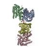
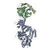
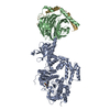
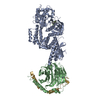
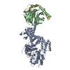
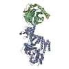
| Title | Crystal structure of the arabidopsis thaliana Raptor in complex with the TOS peptide of human S6K1 | ||||||
 Components Components |
| ||||||
 Keywords Keywords | PROTEIN BINDING / Raptor / TOS | ||||||
| Function / homology |  Function and homology information Function and homology informationmaintenance of shoot apical meristem identity / long-chain fatty acid import into cell / embryo development ending in seed dormancy / negative regulation of TORC2 signaling / TORC1 complex / cellular response to nutrient / Cul4-RING E3 ubiquitin ligase complex / phosphatidylinositol-mediated signaling / germ cell development / TORC1 signaling ...maintenance of shoot apical meristem identity / long-chain fatty acid import into cell / embryo development ending in seed dormancy / negative regulation of TORC2 signaling / TORC1 complex / cellular response to nutrient / Cul4-RING E3 ubiquitin ligase complex / phosphatidylinositol-mediated signaling / germ cell development / TORC1 signaling / TOR signaling / mTORC1-mediated signalling / cellular response to dexamethasone stimulus / positive regulation of translational initiation / behavioral fear response / protein serine/threonine/tyrosine kinase activity / negative regulation of insulin receptor signaling pathway / positive regulation of TORC1 signaling / positive regulation of mitotic cell cycle / cellular response to starvation / peptidyl-serine phosphorylation / negative regulation of autophagy / negative regulation of extrinsic apoptotic signaling pathway / regulation of autophagy / positive regulation of translation / cellular response to amino acid stimulus / G1/S transition of mitotic cell cycle / response to nutrient levels / cellular response to growth factor stimulus / cellular response to type II interferon / modulation of chemical synaptic transmission / kinase binding / cellular response to insulin stimulus / positive regulation of cell growth / protein-macromolecule adaptor activity / protein kinase activity / mitochondrial outer membrane / non-specific serine/threonine protein kinase / neuron projection / protein serine kinase activity / protein serine/threonine kinase activity / apoptotic process / negative regulation of apoptotic process / glutamatergic synapse / signal transduction / mitochondrion / nucleoplasm / ATP binding / nucleus / cytoplasm / cytosol Similarity search - Function | ||||||
| Biological species |   Homo sapiens (human) Homo sapiens (human) | ||||||
| Method |  X-RAY DIFFRACTION / SYNCHROTRON / MOLECULAR REPLACEMENT / Resolution: 3.11 Å X-RAY DIFFRACTION / SYNCHROTRON / MOLECULAR REPLACEMENT / Resolution: 3.11 Å | ||||||
 Authors Authors | Pavletich, N.P. / Jiang, X. | ||||||
 Citation Citation | Journal: Nature / Year: 2017 Title: Mechanisms of mTORC1 activation by RHEB and inhibition by PRAS40. Authors: Haijuan Yang / Xiaolu Jiang / Buren Li / Hyo J Yang / Meredith Miller / Angela Yang / Ankita Dhar / Nikola P Pavletich /  Abstract: The mechanistic target of rapamycin complex 1 (mTORC1) controls cell growth and metabolism in response to nutrients, energy levels, and growth factors. It contains the atypical kinase mTOR and the ...The mechanistic target of rapamycin complex 1 (mTORC1) controls cell growth and metabolism in response to nutrients, energy levels, and growth factors. It contains the atypical kinase mTOR and the RAPTOR subunit that binds to the Tor signalling sequence (TOS) motif of substrates and regulators. mTORC1 is activated by the small GTPase RHEB (Ras homologue enriched in brain) and inhibited by PRAS40. Here we present the 3.0 ångström cryo-electron microscopy structure of mTORC1 and the 3.4 ångström structure of activated RHEB-mTORC1. RHEB binds to mTOR distally from the kinase active site, yet causes a global conformational change that allosterically realigns active-site residues, accelerating catalysis. Cancer-associated hyperactivating mutations map to structural elements that maintain the inactive state, and we provide biochemical evidence that they mimic RHEB relieving auto-inhibition. We also present crystal structures of RAPTOR-TOS motif complexes that define the determinants of TOS recognition, of an mTOR FKBP12-rapamycin-binding (FRB) domain-substrate complex that establishes a second substrate-recruitment mechanism, and of a truncated mTOR-PRAS40 complex that reveals PRAS40 inhibits both substrate-recruitment sites. These findings help explain how mTORC1 selects its substrates, how its kinase activity is controlled, and how it is activated by cancer-associated mutations. | ||||||
| History |
|
- Structure visualization
| Structure viewer | Molecule: MolmilJmol/JSmol |
|---|
- Downloads & links
-Download
| PDBx/mmCIF format | 5wbk.cif.gz | 439.4 KB | Display | PDBx/mmCIF format |
|---|---|---|---|---|
| PDB format | pdb5wbk.ent.gz | 359 KB | Display | PDB format |
| PDBx/mmJSON format | 5wbk.json.gz | Tree view | PDBx/mmJSON format | |
| Others |  Other downloads Other downloads |
-Validation report
| Arichive directory | https://data.pdbj.org/pub/pdb/validation_reports/wb/5wbkftp://data.pdbj.org/pub/pdb/validation_reports/wb/5wbk | HTTPS FTP |
|---|
-Related structure data
| Related structure data |  7086C  7087C  5wbhC  5wbiSC  5wbjC  5wblC  5wbuC  5wbyC  6bcuC  6bcxC S: Starting model for refinement C: citing same article ( |
|---|---|
| Similar structure data |


-Links
 PDBj
PDBj


- Assembly
| Deposited unit | 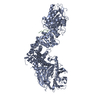
| ||||||||
|---|---|---|---|---|---|---|---|---|---|
| 1 |
| ||||||||
| Unit cell |
|
-Components
| #1: Protein | Mass: 141855.172 Da / Num. of mol.: 1 Source method: isolated from a genetically manipulated source Source: (gene. exp.) Production host: Insect cell expression vector pTIE1 (others) References: UniProt: Q93YQ1 |
|---|---|
| #2: Protein/peptide | Mass: 1564.670 Da / Num. of mol.: 1 / Fragment: residues 24-37 / Source method: obtained synthetically / Source: (synth.) Homo sapiens (human)References: UniProt: P23443, non-specific serine/threonine protein kinase |
-Experimental details
-Experiment
| Experiment | Method: X-RAY DIFFRACTION / Number of used crystals: 1 |
|---|
- Sample preparation
| Crystal | Density Matthews: 2.69 Å3/Da / Density % sol: 54.2 % / Mosaicity: 0.818 ° |
|---|---|
| Crystal grow | Temperature: 289 K / Method: vapor diffusion, hanging drop / pH: 7 / Details: tacsimate |
-Data collection
| Diffraction | Mean temperature: 100 K | ||||||||||||||||||||||||||||||||||||||||||||||||||||||||||||||||||||||||||||||||||||||||||||||||||||||||||||||
|---|---|---|---|---|---|---|---|---|---|---|---|---|---|---|---|---|---|---|---|---|---|---|---|---|---|---|---|---|---|---|---|---|---|---|---|---|---|---|---|---|---|---|---|---|---|---|---|---|---|---|---|---|---|---|---|---|---|---|---|---|---|---|---|---|---|---|---|---|---|---|---|---|---|---|---|---|---|---|---|---|---|---|---|---|---|---|---|---|---|---|---|---|---|---|---|---|---|---|---|---|---|---|---|---|---|---|---|---|---|---|---|
| Diffraction source | Source: SYNCHROTRON / Site: APS / Beamline: 24-ID-C / Wavelength: 0.9792 Å | ||||||||||||||||||||||||||||||||||||||||||||||||||||||||||||||||||||||||||||||||||||||||||||||||||||||||||||||
| Detector | Type: ADSC QUANTUM 315 / Detector: CCD / Date: Feb 12, 2014 | ||||||||||||||||||||||||||||||||||||||||||||||||||||||||||||||||||||||||||||||||||||||||||||||||||||||||||||||
| Radiation | Protocol: SINGLE WAVELENGTH / Monochromatic (M) / Laue (L): M / Scattering type: x-ray | ||||||||||||||||||||||||||||||||||||||||||||||||||||||||||||||||||||||||||||||||||||||||||||||||||||||||||||||
| Radiation wavelength | Wavelength: 0.9792 Å / Relative weight: 1 | ||||||||||||||||||||||||||||||||||||||||||||||||||||||||||||||||||||||||||||||||||||||||||||||||||||||||||||||
| Reflection | Resolution: 3.1→80 Å / Num. obs: 27998 / % possible obs: 99.2 % / Redundancy: 6.5 % / Rmerge(I) obs: 0.084 / Rpim(I) all: 0.035 / Rrim(I) all: 0.091 / Χ2: 0.408 / Net I/σ(I): 5.3 / Num. measured all: 182755 | ||||||||||||||||||||||||||||||||||||||||||||||||||||||||||||||||||||||||||||||||||||||||||||||||||||||||||||||
| Reflection shell | Diffraction-ID: 1
|
- Processing
| Software |
| ||||||||||||||||||||||||||||||||||||||||||||||||||||||||||||||||||||||||||||||||||||||||||||||||||||||||||||||||||||||||||||||||||||||||||||||||||||||||||||||||||||||||||||||||||||||
|---|---|---|---|---|---|---|---|---|---|---|---|---|---|---|---|---|---|---|---|---|---|---|---|---|---|---|---|---|---|---|---|---|---|---|---|---|---|---|---|---|---|---|---|---|---|---|---|---|---|---|---|---|---|---|---|---|---|---|---|---|---|---|---|---|---|---|---|---|---|---|---|---|---|---|---|---|---|---|---|---|---|---|---|---|---|---|---|---|---|---|---|---|---|---|---|---|---|---|---|---|---|---|---|---|---|---|---|---|---|---|---|---|---|---|---|---|---|---|---|---|---|---|---|---|---|---|---|---|---|---|---|---|---|---|---|---|---|---|---|---|---|---|---|---|---|---|---|---|---|---|---|---|---|---|---|---|---|---|---|---|---|---|---|---|---|---|---|---|---|---|---|---|---|---|---|---|---|---|---|---|---|---|---|
| Refinement | Method to determine structure: MOLECULAR REPLACEMENT Starting model: 5WBI Resolution: 3.11→20 Å / Cor.coef. Fo:Fc: 0.944 / Cor.coef. Fo:Fc free: 0.911 / SU B: 55.101 / SU ML: 0.401 / Cross valid method: THROUGHOUT / ESU R Free: 0.477 / Stereochemistry target values: MAXIMUM LIKELIHOOD / Details: HYDROGENS HAVE BEEN ADDED IN THE RIDING POSITIONS
| ||||||||||||||||||||||||||||||||||||||||||||||||||||||||||||||||||||||||||||||||||||||||||||||||||||||||||||||||||||||||||||||||||||||||||||||||||||||||||||||||||||||||||||||||||||||
| Solvent computation | Ion probe radii: 0.9 Å / Shrinkage radii: 0.9 Å / VDW probe radii: 1 Å / Solvent model: MASK | ||||||||||||||||||||||||||||||||||||||||||||||||||||||||||||||||||||||||||||||||||||||||||||||||||||||||||||||||||||||||||||||||||||||||||||||||||||||||||||||||||||||||||||||||||||||
| Displacement parameters | Biso mean: 104.936 Å2
| ||||||||||||||||||||||||||||||||||||||||||||||||||||||||||||||||||||||||||||||||||||||||||||||||||||||||||||||||||||||||||||||||||||||||||||||||||||||||||||||||||||||||||||||||||||||
| Refinement step | Cycle: 1 / Resolution: 3.11→20 Å
| ||||||||||||||||||||||||||||||||||||||||||||||||||||||||||||||||||||||||||||||||||||||||||||||||||||||||||||||||||||||||||||||||||||||||||||||||||||||||||||||||||||||||||||||||||||||
| Refine LS restraints |
|