 Movie
Movie Controller


+ Open data

- Basic information
| Entry | Database: PDB / ID: 4ud7 | |||||||||
|---|---|---|---|---|---|---|---|---|---|---|
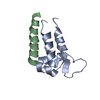
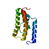
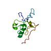
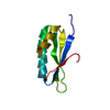
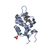
| Title | Structure of the stapled peptide YS-02 bound to MDM2 | |||||||||
 Components Components |
| |||||||||
 Keywords Keywords | LYASE / MDM2 / STAPLED PEPTIDE / P53 | |||||||||
| Function / homology |  Function and homology information Function and homology informationcellular response to vitamin B1 / response to formaldehyde / response to water-immersion restraint stress / response to ether / traversing start control point of mitotic cell cycle / atrial septum development / fibroblast activation / regulation of protein catabolic process at postsynapse, modulating synaptic transmission / Trafficking of AMPA receptors / negative regulation of intrinsic apoptotic signaling pathway by p53 class mediator ...cellular response to vitamin B1 / response to formaldehyde / response to water-immersion restraint stress / response to ether / traversing start control point of mitotic cell cycle / atrial septum development / fibroblast activation / regulation of protein catabolic process at postsynapse, modulating synaptic transmission / Trafficking of AMPA receptors / negative regulation of intrinsic apoptotic signaling pathway by p53 class mediator / receptor serine/threonine kinase binding / negative regulation of protein processing / response to steroid hormone / SUMO transferase activity / peroxisome proliferator activated receptor binding / positive regulation of vascular associated smooth muscle cell migration / atrioventricular valve morphogenesis / AKT phosphorylates targets in the cytosol / response to iron ion / NEDD8 ligase activity / endocardial cushion morphogenesis / ventricular septum development / cellular response to peptide hormone stimulus / positive regulation of muscle cell differentiation / cardiac septum morphogenesis / regulation of postsynaptic neurotransmitter receptor internalization / SUMOylation of ubiquitinylation proteins / cellular response to alkaloid / blood vessel development / ligase activity / Constitutive Signaling by AKT1 E17K in Cancer / negative regulation of DNA damage response, signal transduction by p53 class mediator / cellular response to antibiotic / SUMOylation of transcription factors / negative regulation of signal transduction by p53 class mediator / regulation of protein catabolic process / cellular response to UV-C / cellular response to estrogen stimulus / response to magnesium ion / protein sumoylation / blood vessel remodeling / ribonucleoprotein complex binding / protein localization to nucleus / protein autoubiquitination / positive regulation of vascular associated smooth muscle cell proliferation / NPAS4 regulates expression of target genes / transcription repressor complex / positive regulation of mitotic cell cycle / regulation of heart rate / : / positive regulation of protein export from nucleus / response to cocaine / ubiquitin binding / DNA damage response, signal transduction by p53 class mediator / establishment of protein localization / sperm end piece / Stabilization of p53 / cellular response to gamma radiation / Regulation of RUNX3 expression and activity / RING-type E3 ubiquitin transferase / Oncogene Induced Senescence / Regulation of TP53 Activity through Methylation / protein destabilization / cellular response to growth factor stimulus / Degradation of CDH1 / response to toxic substance / cellular response to hydrogen peroxide / centriolar satellite / disordered domain specific binding / protein polyubiquitination / p53 binding / ubiquitin-protein transferase activity / endocytic vesicle membrane / Signaling by ALK fusions and activated point mutants / Regulation of TP53 Degradation / ubiquitin protein ligase activity / positive regulation of proteasomal ubiquitin-dependent protein catabolic process / negative regulation of neuron projection development / sperm principal piece / 5S rRNA binding / protein-containing complex assembly / sperm midpiece / Oxidative Stress Induced Senescence / cellular response to hypoxia / Regulation of TP53 Activity through Phosphorylation / amyloid fibril formation / ubiquitin-dependent protein catabolic process / proteasome-mediated ubiquitin-dependent protein catabolic process / regulation of cell cycle / postsynaptic density / Ub-specific processing proteases / protein ubiquitination / response to xenobiotic stimulus / protein domain specific binding / response to antibiotic / negative regulation of DNA-templated transcription / apoptotic process / positive regulation of cell population proliferation / ubiquitin protein ligase binding / positive regulation of gene expression Similarity search - Function | |||||||||
| Biological species |  HOMO SAPIENS (human) HOMO SAPIENS (human)SYNTHETIC CONSTRUCT (others) | |||||||||
| Method |  X-RAY DIFFRACTION / SYNCHROTRON / MOLECULAR REPLACEMENT / Resolution: 1.6 Å X-RAY DIFFRACTION / SYNCHROTRON / MOLECULAR REPLACEMENT / Resolution: 1.6 Å | |||||||||
 Authors Authors | Tan, Y.S. / Reeks, J. / Brown, C.J. / Jennings, C.E. / Eapen, R.S. / Tng, Q.S. / Thean, D. / Ying, Y.T. / Gago, F.J.F. / Lane, D.P. ...Tan, Y.S. / Reeks, J. / Brown, C.J. / Jennings, C.E. / Eapen, R.S. / Tng, Q.S. / Thean, D. / Ying, Y.T. / Gago, F.J.F. / Lane, D.P. / Noble, M.E.M. / Verma, C. | |||||||||
 Citation Citation | Journal: J Phys Chem Lett / Year: 2016 Title: Benzene Probes in Molecular Dynamics Simulations Reveal Novel Binding Sites for Ligand Design. Authors: Tan, Y.S. / Reeks, J. / Brown, C.J. / Thean, D. / Ferrer Gago, F.J. / Yuen, T.Y. / Goh, E.T. / Lee, X.E. / Jennings, C.E. / Joseph, T.L. / Lakshminarayanan, R. / Lane, D.P. / Noble, M.E. / Verma, C.S. | |||||||||
| History |
|
- Structure visualization
| Structure viewer | Molecule: MolmilJmol/JSmol |
|---|
- Downloads & links
-Download
| PDBx/mmCIF format | 4ud7.cif.gz | 209.3 KB | Display | PDBx/mmCIF format |
|---|---|---|---|---|
| PDB format | pdb4ud7.ent.gz | 168.1 KB | Display | PDB format |
| PDBx/mmJSON format | 4ud7.json.gz | Tree view | PDBx/mmJSON format | |
| Others |  Other downloads Other downloads |
-Validation report
| Arichive directory | https://data.pdbj.org/pub/pdb/validation_reports/ud/4ud7ftp://data.pdbj.org/pub/pdb/validation_reports/ud/4ud7 | HTTPS FTP |
|---|
-Related structure data
| Related structure data |  4ue1C  1ycrS C: citing same article ( S: Starting model for refinement |
|---|---|
| Similar structure data |


-Links
 PDBj
PDBj


- Assembly
| Deposited unit | 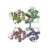
| ||||||||||||||||||||||||||||||||||||||||||||||||||||||||||||||||||||||||||||||||||||||||||||||||||||||||||||||||||||||||||||||||||||||||||||||||||||||
|---|---|---|---|---|---|---|---|---|---|---|---|---|---|---|---|---|---|---|---|---|---|---|---|---|---|---|---|---|---|---|---|---|---|---|---|---|---|---|---|---|---|---|---|---|---|---|---|---|---|---|---|---|---|---|---|---|---|---|---|---|---|---|---|---|---|---|---|---|---|---|---|---|---|---|---|---|---|---|---|---|---|---|---|---|---|---|---|---|---|---|---|---|---|---|---|---|---|---|---|---|---|---|---|---|---|---|---|---|---|---|---|---|---|---|---|---|---|---|---|---|---|---|---|---|---|---|---|---|---|---|---|---|---|---|---|---|---|---|---|---|---|---|---|---|---|---|---|---|---|---|---|
| 1 | 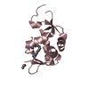
| ||||||||||||||||||||||||||||||||||||||||||||||||||||||||||||||||||||||||||||||||||||||||||||||||||||||||||||||||||||||||||||||||||||||||||||||||||||||
| 2 | 
| ||||||||||||||||||||||||||||||||||||||||||||||||||||||||||||||||||||||||||||||||||||||||||||||||||||||||||||||||||||||||||||||||||||||||||||||||||||||
| 3 | 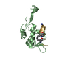
| ||||||||||||||||||||||||||||||||||||||||||||||||||||||||||||||||||||||||||||||||||||||||||||||||||||||||||||||||||||||||||||||||||||||||||||||||||||||
| 4 | 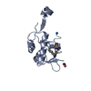
| ||||||||||||||||||||||||||||||||||||||||||||||||||||||||||||||||||||||||||||||||||||||||||||||||||||||||||||||||||||||||||||||||||||||||||||||||||||||
| Unit cell |
| ||||||||||||||||||||||||||||||||||||||||||||||||||||||||||||||||||||||||||||||||||||||||||||||||||||||||||||||||||||||||||||||||||||||||||||||||||||||
| Noncrystallographic symmetry (NCS) | NCS domain:
NCS domain segments: Component-ID: _ / Beg auth comp-ID: SER / Beg label comp-ID: SER / Refine code: _
NCS ensembles :
|
-Components
| #1: Protein | Mass: 12830.667 Da / Num. of mol.: 4 / Fragment: P53 BINDING DOMAIN, UNP RESIDUES 17-125 / Mutation: YES Source method: isolated from a genetically manipulated source Source: (gene. exp.) HOMO SAPIENS (human) / Plasmid: PGEX6P-1 / Production host:  References: UniProt: Q00987, Ligases; Forming carbon-nitrogen bonds; Acid-amino-acid ligases (peptide synthases) #2: Protein/peptide | Mass: 1840.102 Da / Num. of mol.: 4 / Source method: obtained synthetically Details: STAPLED PEPTIDE, COVALENT BOND BETWEEN THE SIDE CHAINS OF RESIDUES 20 AND 24. Source: (synth.) SYNTHETIC CONSTRUCT (others) #3: Water | ChemComp-HOH / |  Mass: 18.015 Da / Num. of mol.: 262 / Source method: isolated from a natural source / Formula: H2O Mass: 18.015 Da / Num. of mol.: 262 / Source method: isolated from a natural source / Formula: H2ONonpolymer details | AMINO GROUP (NH2): STAPLED PEPTIDE IS AMIDATED AT THE C-TERMINUS. ACETYL GROUP (ACE): STAPLED ...AMINO GROUP (NH2): STAPLED PEPTIDE IS AMIDATED AT THE C-TERMINUS. ACETYL GROUP (ACE): STAPLED PEPTIDE IS ACETYLATED | Sequence details | E69 AND K70 ARE MUTATED TO ALANINES FOR SURFACE ENTROPY REDUCTION. THE GPLGS OF THE CRYSTALLISATION ...E69 AND K70 ARE MUTATED TO ALANINES FOR SURFACE ENTROPY REDUCTION. THE GPLGS OF THE CRYSTALLIS | |
|---|
-Experimental details
-Experiment
| Experiment | Method: X-RAY DIFFRACTION / Number of used crystals: 1 |
|---|
- Sample preparation
| Crystal | Density Matthews: 2.8 Å3/Da / Density % sol: 56 % / Description: NONE |
|---|---|
| Crystal grow | pH: 4 / Details: 0.1 M SODIUM CITRATE PH 4.0 AND 15 % PEG8000 |
-Data collection
| Diffraction | Mean temperature: 100 K |
|---|---|
| Diffraction source | Source: SYNCHROTRON / Site: Diamond  / Beamline: I04-1 / Wavelength: 0.92 / Beamline: I04-1 / Wavelength: 0.92 |
| Detector | Type: DECTRIS PILATUS 6M / Detector: PIXEL / Date: Jul 3, 2014 |
| Radiation | Protocol: SINGLE WAVELENGTH / Monochromatic (M) / Laue (L): M / Scattering type: x-ray |
| Radiation wavelength | Wavelength: 0.92 Å / Relative weight: 1 |
| Reflection | Resolution: 1.6→52 Å / Num. obs: 62522 / % possible obs: 97.9 % / Observed criterion σ(I): 1.9 / Redundancy: 3.7 % / Rmerge(I) obs: 0.06 / Net I/σ(I): 12.1 |
| Reflection shell | Resolution: 1.6→1.63 Å / Redundancy: 3.3 % / Rmerge(I) obs: 0.69 / Mean I/σ(I) obs: 1.9 / % possible all: 93.9 |
- Processing
| Software |
| ||||||||||||||||||||||||||||||||||||||||||||||||||||||||||||||||||||||||||||||||||||||||||||||||||||||||||||||||||||||||||||||||||||||||||||||||||||||||||||||||||||||||||||||||||||||
|---|---|---|---|---|---|---|---|---|---|---|---|---|---|---|---|---|---|---|---|---|---|---|---|---|---|---|---|---|---|---|---|---|---|---|---|---|---|---|---|---|---|---|---|---|---|---|---|---|---|---|---|---|---|---|---|---|---|---|---|---|---|---|---|---|---|---|---|---|---|---|---|---|---|---|---|---|---|---|---|---|---|---|---|---|---|---|---|---|---|---|---|---|---|---|---|---|---|---|---|---|---|---|---|---|---|---|---|---|---|---|---|---|---|---|---|---|---|---|---|---|---|---|---|---|---|---|---|---|---|---|---|---|---|---|---|---|---|---|---|---|---|---|---|---|---|---|---|---|---|---|---|---|---|---|---|---|---|---|---|---|---|---|---|---|---|---|---|---|---|---|---|---|---|---|---|---|---|---|---|---|---|---|---|
| Refinement | Method to determine structure: MOLECULAR REPLACEMENT Starting model: PDB ENTRY 1YCR Resolution: 1.6→76.65 Å / Cor.coef. Fo:Fc: 0.977 / Cor.coef. Fo:Fc free: 0.967 / SU B: 3.544 / SU ML: 0.054 / Cross valid method: THROUGHOUT / ESU R: 0.094 / ESU R Free: 0.076 / Stereochemistry target values: MAXIMUM LIKELIHOOD / Details: U VALUES REFINED INDIVIDUALLY
| ||||||||||||||||||||||||||||||||||||||||||||||||||||||||||||||||||||||||||||||||||||||||||||||||||||||||||||||||||||||||||||||||||||||||||||||||||||||||||||||||||||||||||||||||||||||
| Solvent computation | Ion probe radii: 0.8 Å / Shrinkage radii: 0.8 Å / VDW probe radii: 1.2 Å / Solvent model: MASK | ||||||||||||||||||||||||||||||||||||||||||||||||||||||||||||||||||||||||||||||||||||||||||||||||||||||||||||||||||||||||||||||||||||||||||||||||||||||||||||||||||||||||||||||||||||||
| Displacement parameters | Biso mean: 23.018 Å2
| ||||||||||||||||||||||||||||||||||||||||||||||||||||||||||||||||||||||||||||||||||||||||||||||||||||||||||||||||||||||||||||||||||||||||||||||||||||||||||||||||||||||||||||||||||||||
| Refinement step | Cycle: LAST / Resolution: 1.6→76.65 Å
| ||||||||||||||||||||||||||||||||||||||||||||||||||||||||||||||||||||||||||||||||||||||||||||||||||||||||||||||||||||||||||||||||||||||||||||||||||||||||||||||||||||||||||||||||||||||
| Refine LS restraints |
|