 Movie
Movie Controller

[English] 日本語
 Yorodumi
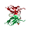
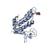
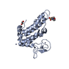
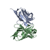
- PDB-2imn: Refined crystal structure of a recombinant immunoglobulin domain ... -
+ Open data

- Basic information
| Entry | Database: PDB / ID: 2imn | ||||||
|---|---|---|---|---|---|---|---|
| Title | Refined crystal structure of a recombinant immunoglobulin domain and a complementarity-determining region 1-grafted mutant | ||||||
 Components Components | IGA-KAPPA MCPC603 FV (LIGHT CHAIN) | ||||||
 Keywords Keywords | IMMUNOGLOBULIN | ||||||
| Function / homology | Immunoglobulins / Immunoglobulin-like / Sandwich / Mainly Beta / ACETATE ION / :  Function and homology information Function and homology information | ||||||
| Biological species |  | ||||||
| Method |  X-RAY DIFFRACTION / Resolution: 1.97 Å X-RAY DIFFRACTION / Resolution: 1.97 Å | ||||||
 Authors Authors | Steipe, B. / Huber, R. | ||||||
 Citation Citation | Journal: J.Mol.Biol. / Year: 1992 Title: Refined crystal structure of a recombinant immunoglobulin domain and a complementarity-determining region 1-grafted mutant. Authors: Steipe, B. / Pluckthun, A. / Huber, R. | ||||||
| History |
|
- Structure visualization
| Structure viewer | Molecule: MolmilJmol/JSmol |
|---|
- Downloads & links
-Download
| PDBx/mmCIF format | 2imn.cif.gz | 37.9 KB | Display | PDBx/mmCIF format |
|---|---|---|---|---|
| PDB format | pdb2imn.ent.gz | 25.4 KB | Display | PDB format |
| PDBx/mmJSON format | 2imn.json.gz | Tree view | PDBx/mmJSON format | |
| Others |  Other downloads Other downloads |
-Validation report
| Arichive directory | https://data.pdbj.org/pub/pdb/validation_reports/im/2imnftp://data.pdbj.org/pub/pdb/validation_reports/im/2imn | HTTPS FTP |
|---|
-Related structure data


-Links
 PDBj
PDBj

- Assembly
| Deposited unit | 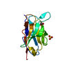
| ||||||||
|---|---|---|---|---|---|---|---|---|---|
| 1 | 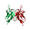
| ||||||||
| 2 | 
| ||||||||
| Unit cell |
| ||||||||
| Atom site foot note | 1: RESIDUES PRO 8 AND PRO 95 ARE CIS-PROLINES. 2: RESIDUES TYR 31, LYS 31A, LYS 103, AND ARG 108 ARE PARTIALLY DISORDERED IN THE ELECTRON DENSITY. | ||||||||
| Components on special symmetry positions |
|
-Components
| #1: Antibody | Mass: 12305.757 Da / Num. of mol.: 1 Source method: isolated from a genetically manipulated source Source: (gene. exp.) |
|---|---|
| #2: Chemical | ChemComp-SO4 /   Mass: 96.063 Da / Num. of mol.: 1 / Source method: obtained synthetically / Formula: SO4 Mass: 96.063 Da / Num. of mol.: 1 / Source method: obtained synthetically / Formula: SO4 |
| #3: Chemical | ChemComp-ACT /   Mass: 59.044 Da / Num. of mol.: 1 / Source method: obtained synthetically / Formula: C2H3O2 Mass: 59.044 Da / Num. of mol.: 1 / Source method: obtained synthetically / Formula: C2H3O2 |
| #4: Water | ChemComp-HOH /  Mass: 18.015 Da / Num. of mol.: 120 / Source method: isolated from a natural source / Formula: H2O Mass: 18.015 Da / Num. of mol.: 120 / Source method: isolated from a natural source / Formula: H2O |
| Has protein modification | Y |
-Experimental details
-Experiment
| Experiment | Method: X-RAY DIFFRACTION |
|---|
- Sample preparation
| Crystal | Density Matthews: 3.27 Å3/Da / Density % sol: 62.41 % |
|---|
-Data collection
| Reflection | *PLUS Highest resolution: 1.9 Å / Num. obs: 11262 / Num. measured all: 96963 / Rmerge(I) obs: 0.086 |
|---|
- Processing
| Software | Name: EREF / Classification: refinement | ||||||||||||||||||||||||||||||||||||||||||||||||||||||||||||
|---|---|---|---|---|---|---|---|---|---|---|---|---|---|---|---|---|---|---|---|---|---|---|---|---|---|---|---|---|---|---|---|---|---|---|---|---|---|---|---|---|---|---|---|---|---|---|---|---|---|---|---|---|---|---|---|---|---|---|---|---|---|
| Refinement | Rfactor Rwork: 0.149 / Highest resolution: 1.97 Å Details: RESIDUES TYR 31, LYS 31A, LYS 103, AND ARG 108 ARE PARTIALLY DISORDERED IN THE ELECTRON DENSITY. | ||||||||||||||||||||||||||||||||||||||||||||||||||||||||||||
| Refinement step | Cycle: LAST / Highest resolution: 1.97 Å
| ||||||||||||||||||||||||||||||||||||||||||||||||||||||||||||
| Refine LS restraints |
| ||||||||||||||||||||||||||||||||||||||||||||||||||||||||||||
| Refinement | *PLUS Highest resolution: 1.97 Å / Rfactor obs: 0.149 | ||||||||||||||||||||||||||||||||||||||||||||||||||||||||||||
| Solvent computation | *PLUS | ||||||||||||||||||||||||||||||||||||||||||||||||||||||||||||
| Displacement parameters | *PLUS | ||||||||||||||||||||||||||||||||||||||||||||||||||||||||||||
| Refine LS restraints | *PLUS Type: o_angle_d / Dev ideal: 2.23 |
