 Movie
Movie Controller

[English] 日本語
 Yorodumi
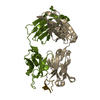
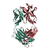
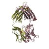
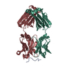
- PDB-2fb4: DIR PRIMAERSTRUKTUR DES KRISTALLISIERBAREN MONOKLONALEN IMMUNOGLO... -
+ Open data

- Basic information
| Entry | Database: PDB / ID: 2fb4 | |||||||||
|---|---|---|---|---|---|---|---|---|---|---|
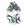
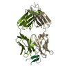
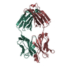
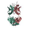
| Title | DIR PRIMAERSTRUKTUR DES KRISTALLISIERBAREN MONOKLONALEN IMMUNOGLOBULINS IGG1 KOL. II. AMINOSAEURESEQUENZ DER L-KETTE, LAMBDA-TYP, SUBGRUPPE I (GERMAN) | |||||||||
 Components Components |
| |||||||||
 Keywords Keywords | IMMUNOGLOBULIN | |||||||||
| Function / homology |  Function and homology information Function and homology informationCD22 mediated BCR regulation / Fc epsilon receptor (FCERI) signaling / Classical antibody-mediated complement activation / IgG immunoglobulin complex / Initial triggering of complement / immunoglobulin mediated immune response / FCGR activation / Role of LAT2/NTAL/LAB on calcium mobilization / immunoglobulin complex / Role of phospholipids in phagocytosis ...CD22 mediated BCR regulation / Fc epsilon receptor (FCERI) signaling / Classical antibody-mediated complement activation / IgG immunoglobulin complex / Initial triggering of complement / immunoglobulin mediated immune response / FCGR activation / Role of LAT2/NTAL/LAB on calcium mobilization / immunoglobulin complex / Role of phospholipids in phagocytosis / Scavenging of heme from plasma / antigen binding / FCERI mediated Ca+2 mobilization / FCGR3A-mediated IL10 synthesis / Regulation of Complement cascade / Antigen activates B Cell Receptor (BCR) leading to generation of second messengers / B cell receptor signaling pathway / Cell surface interactions at the vascular wall / FCGR3A-mediated phagocytosis / FCERI mediated MAPK activation / Regulation of actin dynamics for phagocytic cup formation / Immunoregulatory interactions between a Lymphoid and a non-Lymphoid cell / FCERI mediated NF-kB activation / blood microparticle / Potential therapeutics for SARS / adaptive immune response / immune response / : / extracellular exosome / extracellular region / plasma membrane Similarity search - Function | |||||||||
| Biological species |  Homo sapiens (human) Homo sapiens (human) | |||||||||
| Method |  X-RAY DIFFRACTION / Resolution: 1.9 Å X-RAY DIFFRACTION / Resolution: 1.9 Å | |||||||||
 Authors Authors | Marquart, M. / Huber, R. | |||||||||
 Citation Citation | Journal: Biol.Chem.Hoppe-Seyler / Year: 1989 Title: The primary structure of crystallizable monoclonal immunoglobulin IgG1 Kol. II. Amino acid sequence of the L-chain, gamma-type, subgroup I Authors: Kratzin, H.D. / Palm, W. / Stangel, M. / Schmidt, W.E. / Friedrich, J. / Hilschmann, N. #1: Journal: Immunol.Today / Year: 1982Title: The Three-Dimensional Structure of Antibodies Authors: Marquart, M. / Deisenhofer, J. #2: Journal: J.Mol.Biol. / Year: 1980Title: Crystallographic Refinement and Atomic Models of the Intact Immunoglobulin Molecule Kol and its Antigen-Binding Fragment at 3.0 Angstroms and 1.9 Angstroms Resolution Authors: Marquart, M. / Deisenhofer, J. / Huber, R. / Palm, W. #3: Journal: J.Mol.Biol. / Year: 1978Title: Crystal Structure of the Human Fab Fragment Kol and its Comparison with the Intact Kol Molecule Authors: Matsushima, M. / Marquart, M. / Jones, T.A. / Colman, P.M. / Bartels, K. / Huber, R. #4: Journal: Nature / Year: 1976Title: Crystallographic Structure Studies of an Igg Molecule and an Fc Fragment Authors: Huber, R. / Deisenhofer, J. / Colman, P.M. / Matsushima, M. / Palm, W. #5: Journal: J.Mol.Biol. / Year: 1976Title: Structure of the Human Antibody Molecule Kol (Immunoglobulin G1). An Electron Density Map at 5 Angstroms Resolution Authors: Colman, P.M. / Deisenhofer, J. / Huber, R. / Palm, W. | |||||||||
| History |
|
- Structure visualization
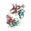
| Structure viewer | Molecule: MolmilJmol/JSmol |
|---|
- Downloads & links
-Download
| PDBx/mmCIF format | 2fb4.cif.gz | 97.6 KB | Display | PDBx/mmCIF format |
|---|---|---|---|---|
| PDB format | pdb2fb4.ent.gz | 74.3 KB | Display | PDB format |
| PDBx/mmJSON format | 2fb4.json.gz | Tree view | PDBx/mmJSON format | |
| Others |  Other downloads Other downloads |
-Validation report
| Arichive directory | https://data.pdbj.org/pub/pdb/validation_reports/fb/2fb4ftp://data.pdbj.org/pub/pdb/validation_reports/fb/2fb4 | HTTPS FTP |
|---|
-Related structure data


-Links
 PDBj
PDBj


- Assembly
| Deposited unit | 
| ||||||||
|---|---|---|---|---|---|---|---|---|---|
| 1 |
| ||||||||
| Unit cell |
| ||||||||
| Atom site foot note | 1: RESIDUES L 143, H 152, H 154 ARE CIS-PROLINES. 2: THESE ATOMS WERE NOT FOUND IN THE ELECTRON DENSITY MAP. THEIR COORDINATES WERE GENERATED USING STEREOCHEMICAL CRITERIA. |
-Components
| #1: Antibody | Mass: 22817.348 Da / Num. of mol.: 1 Source method: isolated from a genetically manipulated source Source: (gene. exp.) Homo sapiens (human) / References: UniProt: P0CG04*PLUS |
|---|---|
| #2: Antibody | Mass: 24325.139 Da / Num. of mol.: 1 Source method: isolated from a genetically manipulated source Source: (gene. exp.) Homo sapiens (human) / References: UniProt: P01772 |
| #3: Water | ChemComp-HOH /  Mass: 18.015 Da / Num. of mol.: 105 / Source method: isolated from a natural source / Formula: H2O Mass: 18.015 Da / Num. of mol.: 105 / Source method: isolated from a natural source / Formula: H2O |
| Has protein modification | Y |
-Experimental details
-Experiment
| Experiment | Method: X-RAY DIFFRACTION |
|---|
- Sample preparation
| Crystal | Density Matthews: 2.31 Å3/Da / Density % sol: 46.65 % |
|---|---|
| Crystal grow | *PLUS Method: unknown |
- Processing
| Software | Name: EREF / Classification: refinement | ||||||||||||
|---|---|---|---|---|---|---|---|---|---|---|---|---|---|
| Refinement | Resolution: 1.9→6 Å / Rfactor Rwork: 0.189 | ||||||||||||
| Refinement step | Cycle: LAST / Resolution: 1.9→6 Å
| ||||||||||||
| Refinement | *PLUS Highest resolution: 1.9 Å / Lowest resolution: 6 Å / Rfactor obs: 0.189 | ||||||||||||
| Solvent computation | *PLUS | ||||||||||||
| Displacement parameters | *PLUS |
