 Movie
Movie Controller

[English] 日本語
 Yorodumi
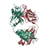
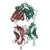
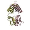
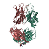
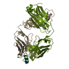
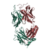
- PDB-1frg: CRYSTAL STRUCTURE, SEQUENCE, AND EPITOPE MAPPING OF A PEPTIDE COM... -
+ Open data

- Basic information
| Entry | Database: PDB / ID: 1frg | ||||||
|---|---|---|---|---|---|---|---|
| Title | CRYSTAL STRUCTURE, SEQUENCE, AND EPITOPE MAPPING OF A PEPTIDE COMPLEX OF AN ANTI-INFLUENZA HA PEPTIDE ANTIBODY FAB 26(SLASH)9: FINE-TUNING ANTIBODY SPECIFICITY | ||||||
 Components Components |
| ||||||
 Keywords Keywords | Viral protein/Immune system / IMMUNOGLOBULIN/VIRUS HEMAGGLUTININ / Viral protein-Immune system COMPLEX | ||||||
| Function / homology |  Function and homology information Function and homology informationalpha-beta T cell receptor complex / immunoglobulin complex, circulating / immunoglobulin receptor binding / IgG immunoglobulin complex / complement activation, classical pathway / antigen binding / B cell differentiation / antibacterial humoral response / adaptive immune response / host cell surface receptor binding ...alpha-beta T cell receptor complex / immunoglobulin complex, circulating / immunoglobulin receptor binding / IgG immunoglobulin complex / complement activation, classical pathway / antigen binding / B cell differentiation / antibacterial humoral response / adaptive immune response / host cell surface receptor binding / fusion of virus membrane with host plasma membrane / fusion of virus membrane with host endosome membrane / viral envelope / symbiont entry into host cell / virion attachment to host cell / host cell plasma membrane / extracellular region / plasma membrane Similarity search - Function | ||||||
| Biological species |  | ||||||
| Method |  X-RAY DIFFRACTION / Resolution: 2.8 Å X-RAY DIFFRACTION / Resolution: 2.8 Å | ||||||
 Authors Authors | Churchill, M.E.A. / Wilson, I.A. | ||||||
 Citation Citation | Journal: J.Mol.Biol. / Year: 1994 Title: Crystal structure of a peptide complex of anti-influenza peptide antibody Fab 26/9. Comparison of two different antibodies bound to the same peptide antigen. Authors: Churchill, M.E. / Stura, E.A. / Pinilla, C. / Appel, J.R. / Houghten, R.A. / Kono, D.H. / Balderas, R.S. / Fieser, G.G. / Schulze-Gahmen, U. / Wilson, I.A. #1: Journal: J.Mol.Biol. / Year: 1993Title: Detailed Analysis of the Free and Bound Conformations of an Antibody: X-Ray Structures of Fab 17(Slash)9 and Three Different Fab-Peptide Complexes Authors: Schulze-Gahmen, U. / Rini, J.M. / Wilson, I.A. #2: Journal: Science / Year: 1992Title: Structural Evidence for Induced Fit as a Mechanism for Antibody-Antigen Recognition Authors: Rini, J.M. / Schulze-Gahmen, U. / Wilson, I.A. #3: Journal: J.Biol.Chem. / Year: 1988Title: Preliminary Crystallographic Data, Primary Sequence, and Binding Data for the Anti-Peptide Fab and its Complex with a Synthetic Peptide from Influenza Hemagglutinin Authors: Schulze-Gahmen, U. / Rini, J.M. / Arevalo, J. / Stura, E.A. / Kenten, J.H. / Wilson, I.A. | ||||||
| History |
|
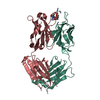
- Structure visualization
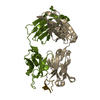
| Structure viewer | Molecule: MolmilJmol/JSmol |
|---|
- Downloads & links
-Download
| PDBx/mmCIF format | 1frg.cif.gz | 99.4 KB | Display | PDBx/mmCIF format |
|---|---|---|---|---|
| PDB format | pdb1frg.ent.gz | 74.8 KB | Display | PDB format |
| PDBx/mmJSON format | 1frg.json.gz | Tree view | PDBx/mmJSON format | |
| Others |  Other downloads Other downloads |
-Validation report
| Arichive directory | https://data.pdbj.org/pub/pdb/validation_reports/fr/1frgftp://data.pdbj.org/pub/pdb/validation_reports/fr/1frg | HTTPS FTP |
|---|
-Related structure data
| Related structure data | |
|---|---|
| Similar structure data |


-Links
 PDBj
PDBj


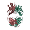
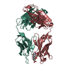
- Assembly
| Deposited unit | 
| ||||||||
|---|---|---|---|---|---|---|---|---|---|
| 1 |
| ||||||||
| Unit cell |
| ||||||||
| Atom site foot note | 1: CIS PROLINE - PRO L 8 / 2: CIS PROLINE - PRO L 101 / 3: CIS PROLINE - PRO L 147 / 4: CIS PROLINE - PRO H 371 / 5: CIS PROLINE - PRO H 373 / 6: CIS PROLINE - PRO H 413 |
-Components
| #1: Antibody | Mass: 24028.650 Da / Num. of mol.: 1 Source method: isolated from a genetically manipulated source Source: (gene. exp.) |
|---|---|
| #2: Antibody | Mass: 23756.715 Da / Num. of mol.: 1 Source method: isolated from a genetically manipulated source Source: (gene. exp.) |
| #3: Protein/peptide | Mass: 902.969 Da / Num. of mol.: 1 Source method: isolated from a genetically manipulated source References: UniProt: E3W6A8*PLUS |
| #4: Water | ChemComp-HOH /  Mass: 18.015 Da / Num. of mol.: 1 / Source method: isolated from a natural source / Formula: H2O Mass: 18.015 Da / Num. of mol.: 1 / Source method: isolated from a natural source / Formula: H2O |
| Has protein modification | Y |
| Nonpolymer details | ONE BOUND WATER IS INCLUDED IN THE PEPTIDE BINDING SITE OF THE FAB. |
| Sequence details | THE LIGHT CHAIN IS NUMBERED AS CHAIN L 1 - 217. THE HEAVY CHAIN IS NUMBERED AS CHAIN H 218 - 437. ...THE LIGHT CHAIN IS NUMBERED AS CHAIN L 1 - 217. THE HEAVY CHAIN IS NUMBERED AS CHAIN H 218 - 437. THE PEPTIDE IS NUMBERED AS CHAIN P 1 - 9. |
-Experimental details
-Experiment
| Experiment | Method: X-RAY DIFFRACTION |
|---|
- Sample preparation
| Crystal | Density Matthews: 2.88 Å3/Da / Density % sol: 57.35 % | |||||||||||||||||||||||||
|---|---|---|---|---|---|---|---|---|---|---|---|---|---|---|---|---|---|---|---|---|---|---|---|---|---|---|
| Crystal grow | *PLUS Temperature: 22.5 ℃ / Method: vapor diffusion, sitting drop / PH range low: 9 / PH range high: 7 | |||||||||||||||||||||||||
| Components of the solutions | *PLUS
|
-Data collection
| Radiation | Scattering type: x-ray |
|---|---|
| Radiation wavelength | Relative weight: 1 |
| Reflection | *PLUS Highest resolution: 2.6 Å / Lowest resolution: 9999 Å / Num. obs: 15290 / % possible obs: 87 % / Num. measured all: 45397 / Rmerge(I) obs: 0.1 |
| Reflection shell | *PLUS Highest resolution: 2.6 Å / Lowest resolution: 2.8 Å / % possible obs: 43 % / Num. unique obs: 1237 / Num. measured obs: 2555 / Rmerge(I) obs: 0.48 |
- Processing
| Software |
| ||||||||||||||||||||||||||||||||||||||||||||||||||||||||||||
|---|---|---|---|---|---|---|---|---|---|---|---|---|---|---|---|---|---|---|---|---|---|---|---|---|---|---|---|---|---|---|---|---|---|---|---|---|---|---|---|---|---|---|---|---|---|---|---|---|---|---|---|---|---|---|---|---|---|---|---|---|---|
| Refinement | Resolution: 2.8→8 Å / Rfactor Rwork: 0.19 / Rfactor obs: 0.19 | ||||||||||||||||||||||||||||||||||||||||||||||||||||||||||||
| Refinement step | Cycle: LAST / Resolution: 2.8→8 Å
| ||||||||||||||||||||||||||||||||||||||||||||||||||||||||||||
| Refine LS restraints |
| ||||||||||||||||||||||||||||||||||||||||||||||||||||||||||||
| Software | *PLUS Name: X-PLOR / Classification: refinement | ||||||||||||||||||||||||||||||||||||||||||||||||||||||||||||
| Refinement | *PLUS Rfactor obs: 0.19 / Rfactor Rwork: 0.19 | ||||||||||||||||||||||||||||||||||||||||||||||||||||||||||||
| Solvent computation | *PLUS | ||||||||||||||||||||||||||||||||||||||||||||||||||||||||||||
| Displacement parameters | *PLUS | ||||||||||||||||||||||||||||||||||||||||||||||||||||||||||||
| Refine LS restraints | *PLUS
|
