 Movie
Movie Controller

[English] 日本語
 Yorodumi
- PDB-1him: STRUCTURAL EVIDENCE FOR INDUCED FIT AS A MECHANISM FOR ANTIBODY-A... -
+ Open data

- Basic information
| Entry | Database: PDB / ID: 1him | ||||||
|---|---|---|---|---|---|---|---|
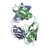
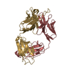
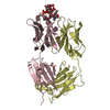
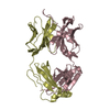
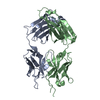
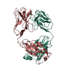
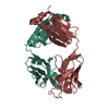
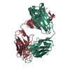
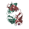
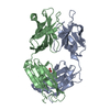
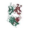
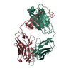
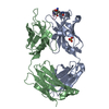
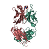
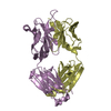
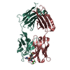
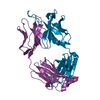
| Title | STRUCTURAL EVIDENCE FOR INDUCED FIT AS A MECHANISM FOR ANTIBODY-ANTIGEN RECOGNITION | ||||||
 Components Components |
| ||||||
 Keywords Keywords | IMMUNOGLOBULIN | ||||||
| Function / homology |  Function and homology information Function and homology informationviral budding from plasma membrane / clathrin-dependent endocytosis of virus by host cell / host cell surface receptor binding / fusion of virus membrane with host plasma membrane / fusion of virus membrane with host endosome membrane / viral envelope / virion attachment to host cell / host cell plasma membrane / virion membrane / membrane Similarity search - Function | ||||||
| Biological species |  | ||||||
| Method |  X-RAY DIFFRACTION / Resolution: 2.9 Å X-RAY DIFFRACTION / Resolution: 2.9 Å | ||||||
 Authors Authors | Schulze-Gahmen, U. / Wilson, I.A. | ||||||
 Citation Citation | Journal: Science / Year: 1992 Title: Structural evidence for induced fit as a mechanism for antibody-antigen recognition. Authors: Rini, J.M. / Schulze-Gahmen, U. / Wilson, I.A. #1: Journal: J.Biol.Chem. / Year: 1988Title: Preliminary Crystallographic Data, Primary Sequence, and Binding Data for an Anti-Peptide Fab and its Complex with a Synthetic Peptide from Influenza Virus Hemagglutinin Authors: Schulze-Gahmen, U. / Rini, J.M. / Arevalo, J. / Stura, E.A. / Kenten, J.H. / Wilson, I.A. | ||||||
| History |
|
- Structure visualization
| Structure viewer | Molecule: MolmilJmol/JSmol |
|---|
- Downloads & links
-Download
| PDBx/mmCIF format | 1him.cif.gz | 174.8 KB | Display | PDBx/mmCIF format |
|---|---|---|---|---|
| PDB format | pdb1him.ent.gz | 137.5 KB | Display | PDB format |
| PDBx/mmJSON format | 1him.json.gz | Tree view | PDBx/mmJSON format | |
| Others |  Other downloads Other downloads |
-Validation report
| Arichive directory | https://data.pdbj.org/pub/pdb/validation_reports/hi/1himftp://data.pdbj.org/pub/pdb/validation_reports/hi/1him | HTTPS FTP |
|---|
-Related structure data


-Links
 PDBj
PDBj


- Assembly
| Deposited unit | 
| ||||||||
|---|---|---|---|---|---|---|---|---|---|
| 1 | 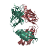
| ||||||||
| 2 | 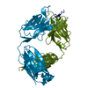
| ||||||||
| Unit cell |
| ||||||||
| Atom site foot note | 1: RESIDUES PRO 8, PRO 95, AND PRO 141 OF LIGHT CHAINS AND RESIDUES PRO 149, PRO 151 AND PRO 200 OF HEAVY CHAINS ARE CIS PROLINES. |
-Components
| #1: Antibody | Mass: 23911.369 Da / Num. of mol.: 2 Source method: isolated from a genetically manipulated source Source: (gene. exp.) #2: Antibody | Mass: 23677.441 Da / Num. of mol.: 2 Source method: isolated from a genetically manipulated source Source: (gene. exp.) #3: Protein/peptide | | Mass: 926.947 Da / Num. of mol.: 1 / Source method: obtained synthetically / References: UniProt: I6SHA6*PLUS #4: Protein/peptide | | Mass: 1040.105 Da / Num. of mol.: 1 / Source method: obtained synthetically / References: UniProt: I6SHA6*PLUS Has protein modification | Y | |
|---|
-Experimental details
-Experiment
| Experiment | Method: X-RAY DIFFRACTION |
|---|
- Sample preparation
| Crystal | Density Matthews: 2.95 Å3/Da / Density % sol: 58.34 % | ||||||||||||||||||||||||
|---|---|---|---|---|---|---|---|---|---|---|---|---|---|---|---|---|---|---|---|---|---|---|---|---|---|
| Crystal grow | *PLUS Method: vapor diffusion, hanging drop / Details: using macroseeding | ||||||||||||||||||||||||
| Components of the solutions | *PLUS
|
-Data collection
| Radiation | Scattering type: x-ray |
|---|---|
| Radiation wavelength | Relative weight: 1 |
| Reflection | *PLUS Highest resolution: 2.9 Å / % possible obs: 87 % / Rmerge(I) obs: 0.12 |
- Processing
| Software |
| ||||||||||||||||||||||||||||||||||||||||||||||||||||||||||||
|---|---|---|---|---|---|---|---|---|---|---|---|---|---|---|---|---|---|---|---|---|---|---|---|---|---|---|---|---|---|---|---|---|---|---|---|---|---|---|---|---|---|---|---|---|---|---|---|---|---|---|---|---|---|---|---|---|---|---|---|---|---|
| Refinement | Resolution: 2.9→8 Å / Rfactor Rwork: 0.2 / Rfactor obs: 0.2 / σ(F): 2 | ||||||||||||||||||||||||||||||||||||||||||||||||||||||||||||
| Refinement step | Cycle: LAST / Resolution: 2.9→8 Å
| ||||||||||||||||||||||||||||||||||||||||||||||||||||||||||||
| Refine LS restraints |
| ||||||||||||||||||||||||||||||||||||||||||||||||||||||||||||
| Software | *PLUS Name: X-PLOR / Classification: refinement | ||||||||||||||||||||||||||||||||||||||||||||||||||||||||||||
| Refinement | *PLUS Highest resolution: 2.9 Å / Lowest resolution: 8 Å / σ(F): 2 / Rfactor obs: 0.2 | ||||||||||||||||||||||||||||||||||||||||||||||||||||||||||||
| Solvent computation | *PLUS | ||||||||||||||||||||||||||||||||||||||||||||||||||||||||||||
| Displacement parameters | *PLUS | ||||||||||||||||||||||||||||||||||||||||||||||||||||||||||||
| Refine LS restraints | *PLUS Type: x_angle_d / Dev ideal: 3.7 |
