 Movie
Movie Controller


+ Open data

- Basic information
| Entry | Database: PDB / ID: 4nuj | ||||||
|---|---|---|---|---|---|---|---|
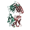
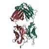
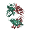
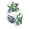
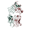
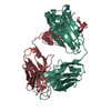
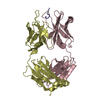
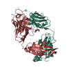
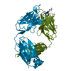
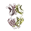
| Title | Crystal structure of HIV-1 broadly neutralizing antibody PGT152 | ||||||
 Components Components |
| ||||||
 Keywords Keywords | IMMUNE SYSTEM / immunoglobulin / Fab fragment / HIV Envelope | ||||||
| Function / homology |  Function and homology information Function and homology informationimmunoglobulin complex / adaptive immune response / extracellular region / metal ion binding / plasma membrane Similarity search - Function | ||||||
| Biological species |  Homo sapiens (human) Homo sapiens (human) | ||||||
| Method |  X-RAY DIFFRACTION / SYNCHROTRON / MOLECULAR REPLACEMENT / Resolution: 1.827 Å X-RAY DIFFRACTION / SYNCHROTRON / MOLECULAR REPLACEMENT / Resolution: 1.827 Å | ||||||
 Authors Authors | Blattner, C. / Wilson, I.A. | ||||||
 Citation Citation | Journal: Immunity / Year: 2014 Title: Structural delineation of a quaternary, cleavage-dependent epitope at the gp41-gp120 interface on intact HIV-1 Env trimers. Authors: Claudia Blattner / Jeong Hyun Lee / Kwinten Sliepen / Ronald Derking / Emilia Falkowska / Alba Torrents de la Peña / Albert Cupo / Jean-Philippe Julien / Marit van Gils / Peter S Lee / ...Authors: Claudia Blattner / Jeong Hyun Lee / Kwinten Sliepen / Ronald Derking / Emilia Falkowska / Alba Torrents de la Peña / Albert Cupo / Jean-Philippe Julien / Marit van Gils / Peter S Lee / Wenjie Peng / James C Paulson / Pascal Poignard / Dennis R Burton / John P Moore / Rogier W Sanders / Ian A Wilson / Andrew B Ward /   Abstract: All previously characterized broadly neutralizing antibodies to the HIV-1 envelope glycoprotein (Env) target one of four major sites of vulnerability. Here, we define and structurally characterize a ...All previously characterized broadly neutralizing antibodies to the HIV-1 envelope glycoprotein (Env) target one of four major sites of vulnerability. Here, we define and structurally characterize a unique epitope on Env that is recognized by a recently discovered family of human monoclonal antibodies (PGT151-PGT158). The PGT151 epitope is comprised of residues and glycans at the interface of gp41 and gp120 within a single protomer and glycans from both subunits of a second protomer and represents a neutralizing epitope that is dependent on both gp120 and gp41. Because PGT151 binds only to properly formed, cleaved trimers, this distinctive property, and its ability to stabilize Env trimers, has enabled the successful purification of mature, cleaved Env trimers from the cell surface as a complex with PGT151. Here we compare the structural and functional properties of membrane-extracted Env trimers from several clades with those of the soluble, cleaved SOSIP gp140 trimer. | ||||||
| History |
|
- Structure visualization
| Structure viewer | Molecule: MolmilJmol/JSmol |
|---|
- Downloads & links
-Download
| PDBx/mmCIF format | 4nuj.cif.gz | 204.2 KB | Display | PDBx/mmCIF format |
|---|---|---|---|---|
| PDB format | pdb4nuj.ent.gz | 163.9 KB | Display | PDB format |
| PDBx/mmJSON format | 4nuj.json.gz | Tree view | PDBx/mmJSON format | |
| Others |  Other downloads Other downloads |
-Validation report
| Arichive directory | https://data.pdbj.org/pub/pdb/validation_reports/nu/4nujftp://data.pdbj.org/pub/pdb/validation_reports/nu/4nuj | HTTPS FTP |
|---|
-Related structure data
| Related structure data |  5918C  5919C  5920C  5921C 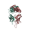 4nugSC S: Starting model for refinement C: citing same article ( |
|---|---|
| Similar structure data |


-Links
 PDBj
PDBj

- Assembly
| Deposited unit | 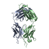
| ||||||||
|---|---|---|---|---|---|---|---|---|---|
| 1 |
| ||||||||
| Unit cell |
| ||||||||
| Components on special symmetry positions |
|
-Components
| #1: Antibody | Mass: 24055.770 Da / Num. of mol.: 1 / Mutation: N107K Source method: isolated from a genetically manipulated source Source: (gene. exp.) Homo sapiens (human) / Cell line (production host): HEK 293F / Production host: Homo sapiens (human) / References: UniProt: Q8TCD0 |
|---|---|
| #2: Antibody | Mass: 26022.338 Da / Num. of mol.: 1 Source method: isolated from a genetically manipulated source Source: (gene. exp.) Homo sapiens (human) / Gene: DKFZp686P15220 / Cell line (production host): HEK 293F / Production host: Homo sapiens (human) / References: UniProt: Q6N089, UniProt: S6BAM6*PLUS |
| #3: Water | ChemComp-HOH /  Mass: 18.015 Da / Num. of mol.: 512 / Source method: isolated from a natural source / Formula: H2O Mass: 18.015 Da / Num. of mol.: 512 / Source method: isolated from a natural source / Formula: H2O |
| Has protein modification | Y |
-Experimental details
-Experiment
| Experiment | Method: X-RAY DIFFRACTION / Number of used crystals: 1 |
|---|
- Sample preparation
| Crystal | Density Matthews: 2.42 Å3/Da / Density % sol: 49.17 % |
|---|---|
| Crystal grow | Temperature: 293.15 K / Method: vapor diffusion / pH: 4.2 Details: 40% PEG 600, 0.1M phosphate-citrate, pH 4.2, VAPOR DIFFUSION, temperature 293.15K |
-Data collection
| Diffraction | Mean temperature: 100 K |
|---|---|
| Diffraction source | Source: SYNCHROTRON / Site: APS / Beamline: 23-ID-B / Wavelength: 1.0332 Å |
| Detector | Type: MARMOSAIC 300 mm CCD / Detector: CCD / Date: Nov 17, 2012 |
| Radiation | Monochromator: Double crystal cryo-cooled Si(111) / Protocol: SINGLE WAVELENGTH / Monochromatic (M) / Laue (L): M / Scattering type: x-ray |
| Radiation wavelength | Wavelength: 1.0332 Å / Relative weight: 1 |
| Reflection | Resolution: 1.8→33 Å / Num. all: 42250 / Num. obs: 42208 / % possible obs: 99.9 % / Observed criterion σ(F): 0 / Observed criterion σ(I): 0 / Redundancy: 7.4 % / Biso Wilson estimate: 22.8 Å2 / Rsym value: 0.08 / Net I/σ(I): 28.9 |
- Processing
| Software |
| ||||||||||||||||||||||||||||||||||||||||||||||||||||||||||||||||||||||||||||||||||||||||||||||||||||||||||||||||
|---|---|---|---|---|---|---|---|---|---|---|---|---|---|---|---|---|---|---|---|---|---|---|---|---|---|---|---|---|---|---|---|---|---|---|---|---|---|---|---|---|---|---|---|---|---|---|---|---|---|---|---|---|---|---|---|---|---|---|---|---|---|---|---|---|---|---|---|---|---|---|---|---|---|---|---|---|---|---|---|---|---|---|---|---|---|---|---|---|---|---|---|---|---|---|---|---|---|---|---|---|---|---|---|---|---|---|---|---|---|---|---|---|---|
| Refinement | Method to determine structure: MOLECULAR REPLACEMENT Starting model: PDB ENTRY 4NUG Resolution: 1.827→32.859 Å / SU ML: 0.19 / σ(F): 1.34 / Phase error: 22.38 / Stereochemistry target values: ML
| ||||||||||||||||||||||||||||||||||||||||||||||||||||||||||||||||||||||||||||||||||||||||||||||||||||||||||||||||
| Solvent computation | Shrinkage radii: 0.9 Å / VDW probe radii: 1.11 Å / Solvent model: FLAT BULK SOLVENT MODEL | ||||||||||||||||||||||||||||||||||||||||||||||||||||||||||||||||||||||||||||||||||||||||||||||||||||||||||||||||
| Refinement step | Cycle: LAST / Resolution: 1.827→32.859 Å
| ||||||||||||||||||||||||||||||||||||||||||||||||||||||||||||||||||||||||||||||||||||||||||||||||||||||||||||||||
| Refine LS restraints |
| ||||||||||||||||||||||||||||||||||||||||||||||||||||||||||||||||||||||||||||||||||||||||||||||||||||||||||||||||
| LS refinement shell |
| ||||||||||||||||||||||||||||||||||||||||||||||||||||||||||||||||||||||||||||||||||||||||||||||||||||||||||||||||
| Refinement TLS params. | Method: refined / Origin x: 18.0616 Å / Origin y: -22.719 Å / Origin z: 15.5145 Å
| ||||||||||||||||||||||||||||||||||||||||||||||||||||||||||||||||||||||||||||||||||||||||||||||||||||||||||||||||
| Refinement TLS group | Selection details: all |
