 Movie
Movie Controller


+ Open data

- Basic information
| Entry | Database: PDB / ID: 2ipu | ||||||
|---|---|---|---|---|---|---|---|
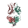
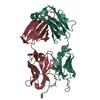
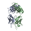
| Title | PFA1 Fab fragment complexed with Abeta 1-8 peptide | ||||||
 Components Components |
| ||||||
 Keywords Keywords | IMMUNE SYSTEM / wwddd / cdr / abeta | ||||||
| Function / homology |  Function and homology information Function and homology informationamyloid-beta complex / growth cone lamellipodium / cellular response to norepinephrine stimulus / collateral sprouting in absence of injury / growth cone filopodium / microglia development / Formyl peptide receptors bind formyl peptides and many other ligands / axo-dendritic transport / regulation of Wnt signaling pathway / axon midline choice point recognition ...amyloid-beta complex / growth cone lamellipodium / cellular response to norepinephrine stimulus / collateral sprouting in absence of injury / growth cone filopodium / microglia development / Formyl peptide receptors bind formyl peptides and many other ligands / axo-dendritic transport / regulation of Wnt signaling pathway / axon midline choice point recognition / regulation of synapse structure or activity / hippocampal neuron apoptotic process / astrocyte activation involved in immune response / NMDA selective glutamate receptor signaling pathway / regulation of spontaneous synaptic transmission / mating behavior / growth factor receptor binding / peptidase activator activity / Insertion of tail-anchored proteins into the endoplasmic reticulum membrane / positive regulation of amyloid fibril formation / PTB domain binding / Golgi-associated vesicle / astrocyte projection / Lysosome Vesicle Biogenesis / Deregulated CDK5 triggers multiple neurodegenerative pathways in Alzheimer's disease models / neuron remodeling / nuclear envelope lumen / TRAF6 mediated NF-kB activation / dendrite development / positive regulation of protein metabolic process / signaling receptor activator activity / negative regulation of long-term synaptic potentiation / Advanced glycosylation endproduct receptor signaling / transition metal ion binding / The NLRP3 inflammasome / modulation of excitatory postsynaptic potential / main axon / intracellular copper ion homeostasis / regulation of multicellular organism growth / ECM proteoglycans / immunoglobulin complex / response to insulin-like growth factor stimulus / positive regulation of T cell migration / regulation of presynapse assembly / neuronal dense core vesicle / Purinergic signaling in leishmaniasis infection / cellular response to manganese ion / positive regulation of chemokine production / Notch signaling pathway / swimming behavior / neuron projection maintenance / extracellular matrix organization / clathrin-coated pit / positive regulation of mitotic cell cycle / axonogenesis / Mitochondrial protein degradation / positive regulation of calcium-mediated signaling / ionotropic glutamate receptor signaling pathway / platelet alpha granule lumen / astrocyte activation / response to interleukin-1 / cellular response to cAMP / regulation of neuron apoptotic process / cellular response to copper ion / positive regulation of glycolytic process / endosome lumen / trans-Golgi network membrane / positive regulation of interleukin-1 beta production / protein serine/threonine kinase binding / dendritic shaft / positive regulation of long-term synaptic potentiation / learning / central nervous system development / Post-translational protein phosphorylation / adult locomotory behavior / serine-type endopeptidase inhibitor activity / locomotory behavior / microglial cell activation / cellular response to nerve growth factor stimulus / TAK1-dependent IKK and NF-kappa-B activation / positive regulation of non-canonical NF-kappaB signal transduction / synapse organization / visual learning / recycling endosome / positive regulation of interleukin-6 production / positive regulation of JNK cascade / regulation of long-term neuronal synaptic plasticity / Golgi lumen / response to lead ion / cognition / Regulation of Insulin-like Growth Factor (IGF) transport and uptake by Insulin-like Growth Factor Binding Proteins (IGFBPs) / cellular response to amyloid-beta / endocytosis / neuron projection development / positive regulation of tumor necrosis factor production / positive regulation of inflammatory response / calcium ion transport / Platelet degranulation / regulation of translation / heparin binding Similarity search - Function | ||||||
| Biological species |  | ||||||
| Method |  X-RAY DIFFRACTION / MOLECULAR REPLACEMENT / Resolution: 1.65 Å X-RAY DIFFRACTION / MOLECULAR REPLACEMENT / Resolution: 1.65 Å | ||||||
 Authors Authors | Gardberg, A.S. / Dealwis, C. | ||||||
 Citation Citation | Journal: Proc.Natl.Acad.Sci.Usa / Year: 2007 Title: Molecular basis for passive immunotherapy of Alzheimer's disease Authors: Gardberg, A.S. / Dice, L.T. / Ou, S. / Rich, R.L. / Helmbrecht, E. / Ko, J. / Wetzel, R. / Myszka, D.G. / Patterson, P.H. / Dealwis, C. | ||||||
| History |
| ||||||
| Remark 999 | SEQUENCE Presently, there is no aminoacid sequence database reference available for the two ...SEQUENCE Presently, there is no aminoacid sequence database reference available for the two proteins IGG2A FAB fragment heavy chain and IGG2A FAB fragment light chain kappa |
- Structure visualization
| Structure viewer | Molecule: MolmilJmol/JSmol |
|---|
- Downloads & links
-Download
| PDBx/mmCIF format | 2ipu.cif.gz | 181.5 KB | Display | PDBx/mmCIF format |
|---|---|---|---|---|
| PDB format | pdb2ipu.ent.gz | 144.5 KB | Display | PDB format |
| PDBx/mmJSON format | 2ipu.json.gz | Tree view | PDBx/mmJSON format | |
| Others |  Other downloads Other downloads |
-Validation report
| Arichive directory | https://data.pdbj.org/pub/pdb/validation_reports/ip/2ipuftp://data.pdbj.org/pub/pdb/validation_reports/ip/2ipu | HTTPS FTP |
|---|
-Related structure data
| Related structure data |  2iptC  2iq9SC  2iqaC  2r0wC 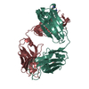 2r0zC  2iqb C: citing same article ( S: Starting model for refinement |
|---|---|
| Similar structure data |


-Links
 PDBj
PDBj


- Assembly
| Deposited unit | 
| ||||||||
|---|---|---|---|---|---|---|---|---|---|
| 1 | 
| ||||||||
| 2 | 
| ||||||||
| Unit cell |
| ||||||||
| Details | The biological assembly is a heterodimer with abeta peptide at the interface. |
-Components
-Protein/peptide , 1 types, 2 molecules PQ
| #3: Protein/peptide | Mass: 977.975 Da / Num. of mol.: 2 / Source method: obtained synthetically Details: This sequence occurs naturally in Homo sapiens (humans) References: UniProt: P05067*PLUS |
|---|
-Antibody , 2 types, 4 molecules LKHG
| #1: Antibody | Mass: 24146.812 Da / Num. of mol.: 2 / Source method: isolated from a natural source / Details: hybridoma / Source: (natural) #2: Antibody | Mass: 24095.115 Da / Num. of mol.: 2 / Source method: isolated from a natural source / Details: hybridoma / Source: (natural) |
|---|
-Non-polymers , 3 types, 127 molecules 


| #4: Chemical |  Mass: 59.067 Da / Num. of mol.: 3 / Source method: obtained synthetically / Formula: C2H5NO Mass: 59.067 Da / Num. of mol.: 3 / Source method: obtained synthetically / Formula: C2H5NO#5: Chemical | ChemComp-GOL /  Mass: 92.094 Da / Num. of mol.: 6 / Source method: obtained synthetically / Formula: C3H8O3 Mass: 92.094 Da / Num. of mol.: 6 / Source method: obtained synthetically / Formula: C3H8O3#6: Water | ChemComp-HOH / | Mass: 18.015 Da / Num. of mol.: 118 / Source method: isolated from a natural source / Formula: H2O |
|---|
-Details
| Has protein modification | Y |
|---|
-Experimental details
-Experiment
| Experiment | Method: X-RAY DIFFRACTION / Number of used crystals: 1 |
|---|
- Sample preparation
| Crystal | Density Matthews: 2.17 Å3/Da / Density % sol: 43.29 % |
|---|---|
| Crystal grow | Temperature: 294 K / Method: vapor diffusion / pH: 8.5 Details: 0.1 M Tris, 25% PEG3350, pH 8.5, VAPOR DIFFUSION, temperature 294K |
-Data collection
| Diffraction | Mean temperature: 113 K |
|---|---|
| Diffraction source | Source: ROTATING ANODE / Type: RIGAKU / Wavelength: 1.54 Å |
| Detector | Type: RIGAKU RAXIS IV / Detector: IMAGE PLATE / Date: Apr 20, 2006 / Details: mirrors |
| Radiation | Protocol: SINGLE WAVELENGTH / Monochromatic (M) / Laue (L): M / Scattering type: x-ray |
| Radiation wavelength | Wavelength: 1.54 Å / Relative weight: 1 |
| Reflection | Resolution: 1.65→23.53 Å / Num. obs: 90408 / % possible obs: 90.7 % / Redundancy: 3.1 % / Rsym value: 0.055 |
| Reflection shell | Resolution: 1.65→1.71 Å / Redundancy: 2.9 % / Mean I/σ(I) obs: 2.4 / Rsym value: 0.467 / % possible all: 66.3 |
- Processing
| Software |
| |||||||||||||||||||||||||||||||||||||||||||||||||||||||||||||||||||||||||||||||||||||||||||||||||||||||||||||||||||||||||||||
|---|---|---|---|---|---|---|---|---|---|---|---|---|---|---|---|---|---|---|---|---|---|---|---|---|---|---|---|---|---|---|---|---|---|---|---|---|---|---|---|---|---|---|---|---|---|---|---|---|---|---|---|---|---|---|---|---|---|---|---|---|---|---|---|---|---|---|---|---|---|---|---|---|---|---|---|---|---|---|---|---|---|---|---|---|---|---|---|---|---|---|---|---|---|---|---|---|---|---|---|---|---|---|---|---|---|---|---|---|---|---|---|---|---|---|---|---|---|---|---|---|---|---|---|---|---|---|
| Refinement | Method to determine structure: MOLECULAR REPLACEMENT Starting model: PDB ENTRY 2IQ9 Resolution: 1.65→23.53 Å / Cor.coef. Fo:Fc: 0.962 / Cor.coef. Fo:Fc free: 0.944 / SU B: 2.106 / SU ML: 0.073 / Cross valid method: THROUGHOUT / σ(F): 0 / ESU R: 0.12 / ESU R Free: 0.118 / Stereochemistry target values: MAXIMUM LIKELIHOOD / Details: HYDROGENS HAVE BEEN ADDED IN THE RIDING POSITIONS
| |||||||||||||||||||||||||||||||||||||||||||||||||||||||||||||||||||||||||||||||||||||||||||||||||||||||||||||||||||||||||||||
| Solvent computation | Ion probe radii: 0.8 Å / Shrinkage radii: 0.8 Å / VDW probe radii: 1.4 Å / Solvent model: MASK | |||||||||||||||||||||||||||||||||||||||||||||||||||||||||||||||||||||||||||||||||||||||||||||||||||||||||||||||||||||||||||||
| Displacement parameters | Biso mean: 14.361 Å2
| |||||||||||||||||||||||||||||||||||||||||||||||||||||||||||||||||||||||||||||||||||||||||||||||||||||||||||||||||||||||||||||
| Refinement step | Cycle: LAST / Resolution: 1.65→23.53 Å
| |||||||||||||||||||||||||||||||||||||||||||||||||||||||||||||||||||||||||||||||||||||||||||||||||||||||||||||||||||||||||||||
| Refine LS restraints |
| |||||||||||||||||||||||||||||||||||||||||||||||||||||||||||||||||||||||||||||||||||||||||||||||||||||||||||||||||||||||||||||
| LS refinement shell | Resolution: 1.65→1.69 Å / Total num. of bins used: 20
|
