 Movie
Movie Controller

[English] 日本語
 Yorodumi
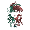
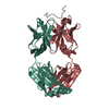
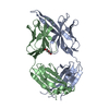
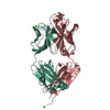
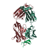
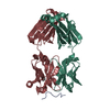
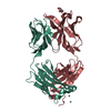
- PDB-3mlt: Crystal structure of anti-HIV-1 V3 Fab 2557 in complex with a UG1... -
+ Open data

- Basic information
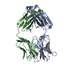
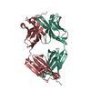
| Entry | Database: PDB / ID: 3mlt | ||||||
|---|---|---|---|---|---|---|---|
| Title | Crystal structure of anti-HIV-1 V3 Fab 2557 in complex with a UG1033 V3 peptide | ||||||
 Components Components |
| ||||||
 Keywords Keywords | IMMUNE SYSTEM / human monoclonal antibody / Fab / HIV-1 / gp120 / third variable loop / antibody-antigen interaction | ||||||
| Function / homology |  Function and homology information Function and homology informationmembrane fusion involved in viral entry into host cell / viral envelope / symbiont entry into host cell / virion attachment to host cell / virion membrane Similarity search - Function | ||||||
| Biological species |  Homo sapiens (human) Homo sapiens (human)  Human immunodeficiency virus 1 Human immunodeficiency virus 1 | ||||||
| Method |  X-RAY DIFFRACTION / SYNCHROTRON / MOLECULAR REPLACEMENT / Resolution: 2.49 Å X-RAY DIFFRACTION / SYNCHROTRON / MOLECULAR REPLACEMENT / Resolution: 2.49 Å | ||||||
 Authors Authors | Kong, X.-P. | ||||||
 Citation Citation | Journal: Nat.Struct.Mol.Biol. / Year: 2010 Title: Conserved structural elements in the V3 crown of HIV-1 gp120. Authors: Jiang, X. / Burke, V. / Totrov, M. / Williams, C. / Cardozo, T. / Gorny, M.K. / Zolla-Pazner, S. / Kong, X.P. | ||||||
| History |
|
- Structure visualization
| Structure viewer | Molecule: MolmilJmol/JSmol |
|---|
- Downloads & links
-Download
| PDBx/mmCIF format | 3mlt.cif.gz | 339.4 KB | Display | PDBx/mmCIF format |
|---|---|---|---|---|
| PDB format | pdb3mlt.ent.gz | 276.2 KB | Display | PDB format |
| PDBx/mmJSON format | 3mlt.json.gz | Tree view | PDBx/mmJSON format | |
| Others |  Other downloads Other downloads |
-Validation report
| Arichive directory | https://data.pdbj.org/pub/pdb/validation_reports/ml/3mltftp://data.pdbj.org/pub/pdb/validation_reports/ml/3mlt | HTTPS FTP |
|---|
-Related structure data
| Related structure data | 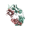 3go1C 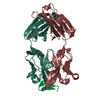 3mlrC 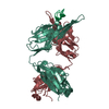 3mlsC 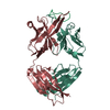 3mluC  3mlvC 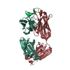 3mlwC 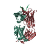 3mlxC 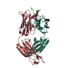 3mlyC 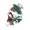 3mlzC C: citing same article ( |
|---|---|
| Similar structure data |
-Links
 PDBj
PDBj


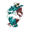
- Assembly
| Deposited unit | 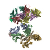
| ||||||||
|---|---|---|---|---|---|---|---|---|---|
| 1 | 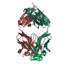
| ||||||||
| 2 | 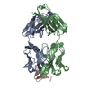
| ||||||||
| 3 | 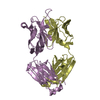
| ||||||||
| 4 | 
| ||||||||
| Unit cell |
|
-Components
| #1: Antibody | Mass: 23331.873 Da / Num. of mol.: 4 / Source method: isolated from a natural source / Source: (natural) Homo sapiens (human)#2: Antibody | Mass: 24191.055 Da / Num. of mol.: 4 / Source method: isolated from a natural source / Source: (natural) Homo sapiens (human)#3: Protein/peptide | Mass: 2462.764 Da / Num. of mol.: 2 / Source method: isolated from a natural source / Source: (natural) Human immunodeficiency virus 1 / References: UniProt: Q9WNX3#4: Water | ChemComp-HOH / |  Mass: 18.015 Da / Num. of mol.: 328 / Source method: isolated from a natural source / Formula: H2O Mass: 18.015 Da / Num. of mol.: 328 / Source method: isolated from a natural source / Formula: H2OHas protein modification | Y | Sequence details | AUTHORS STATE THAT THE FAB WERE MADE BY ENZYME DIGESTION, THEREFORE THE REAL ENDINGS OF THE CHAINS ...AUTHORS STATE THAT THE FAB WERE MADE BY ENZYME DIGESTION, THEREFORE THE REAL ENDINGS OF THE CHAINS ARE UNKNOWN. | |
|---|
-Experimental details
-Experiment
| Experiment | Method: X-RAY DIFFRACTION / Number of used crystals: 1 |
|---|
- Sample preparation
| Crystal | Density Matthews: 2.35 Å3/Da / Density % sol: 47.63 % |
|---|---|
| Crystal grow | Temperature: 296 K / Method: vapor diffusion, hanging drop Details: 20% PEG 3350, 0.2 M Na formate, VAPOR DIFFUSION, HANGING DROP, temperature 296K |
-Data collection
| Diffraction | Mean temperature: 100 K | |||||||||||||||||||||||||||||||||||||||||||||||||||||||
|---|---|---|---|---|---|---|---|---|---|---|---|---|---|---|---|---|---|---|---|---|---|---|---|---|---|---|---|---|---|---|---|---|---|---|---|---|---|---|---|---|---|---|---|---|---|---|---|---|---|---|---|---|---|---|---|---|
| Diffraction source | Source: SYNCHROTRON / Site: NSLS  / Beamline: X4C / Wavelength: 0.9795 Å / Beamline: X4C / Wavelength: 0.9795 Å | |||||||||||||||||||||||||||||||||||||||||||||||||||||||
| Detector | Type: MAR scanner 345 mm plate / Detector: IMAGE PLATE / Date: Jun 11, 2007 Details: Bent single Si (111) crystal monochromator (horizontal focusing and deflection) with vertical focusing mirror | |||||||||||||||||||||||||||||||||||||||||||||||||||||||
| Radiation | Protocol: SINGLE WAVELENGTH / Monochromatic (M) / Laue (L): M / Scattering type: x-ray | |||||||||||||||||||||||||||||||||||||||||||||||||||||||
| Radiation wavelength | Wavelength: 0.9795 Å / Relative weight: 1 | |||||||||||||||||||||||||||||||||||||||||||||||||||||||
| Reflection | Resolution: 2.49→50 Å / Num. obs: 62552 / % possible obs: 99.9 % / Rmerge(I) obs: 0.047 / Χ2: 0.335 / Net I/σ(I): 8.1 | |||||||||||||||||||||||||||||||||||||||||||||||||||||||
| Reflection shell |
|
- Processing
| Software |
| ||||||||||||||||||||||||||||||||||||||||
|---|---|---|---|---|---|---|---|---|---|---|---|---|---|---|---|---|---|---|---|---|---|---|---|---|---|---|---|---|---|---|---|---|---|---|---|---|---|---|---|---|---|
| Refinement | Method to determine structure: MOLECULAR REPLACEMENT / Resolution: 2.49→35.74 Å / Cor.coef. Fo:Fc: 0.938 / Cor.coef. Fo:Fc free: 0.88 / Occupancy max: 1 / Occupancy min: 0.5 / SU B: 11.082 / SU ML: 0.251 / Cross valid method: THROUGHOUT / σ(F): 0 / ESU R: 0.863 / ESU R Free: 0.341 / Stereochemistry target values: MAXIMUM LIKELIHOOD / Details: HYDROGENS HAVE BEEN ADDED IN THE RIDING POSITIONS
| ||||||||||||||||||||||||||||||||||||||||
| Solvent computation | Ion probe radii: 0.8 Å / Shrinkage radii: 0.8 Å / VDW probe radii: 1.2 Å / Solvent model: MASK | ||||||||||||||||||||||||||||||||||||||||
| Displacement parameters | Biso max: 92.51 Å2 / Biso mean: 44.123 Å2 / Biso min: 11.29 Å2
| ||||||||||||||||||||||||||||||||||||||||
| Refinement step | Cycle: LAST / Resolution: 2.49→35.74 Å
| ||||||||||||||||||||||||||||||||||||||||
| LS refinement shell | Resolution: 2.49→2.555 Å / Total num. of bins used: 20
|
