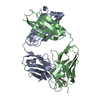
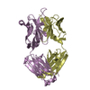
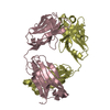
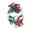
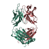
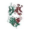
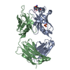
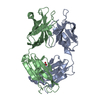
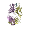
Entry Database : PDB / ID : 4c83Title Crystal Structure of the IgG2a LPT3 in complex with an 8-sugar inner core analogue of Neisseria meningitidis LPT3 HEAVY CHAIN LPT3 LIGHT CHAIN Keywords / / / Function / homology / / / Biological species MUS MUSCULUS (house mouse)Method / / / Resolution : 2.69 Å Authors Parker, M.J. / Gomery, K. / Richard, G. / Mackenzie, C.R. / Cox, A.D. / Richards, J.C. / Evans, S.V. Journal : Glycobiology / Year : 2014Title : Structural Basis for Selective Cross-Reactivity in a Bactericidal Antibody Against Inner Core Lipooligosaccharide from Neisseria Meningitidis.Authors : Parker, M.J. / Gomery, K. / Richard, G. / Mackenzie, C.R. / Cox, A.D. / Richards, J.C. / Evans, S.V. History Deposition Sep 29, 2013 Deposition site / Processing site Revision 1.0 Feb 12, 2014 Provider / Type Revision 1.1 Apr 16, 2014 Group Revision 1.2 Sep 23, 2015 Group Revision 2.0 Jul 29, 2020 Group Atomic model / Data collection ... Atomic model / Data collection / Derived calculations / Other / Structure summary Category atom_site / chem_comp ... atom_site / chem_comp / entity / pdbx_branch_scheme / pdbx_chem_comp_identifier / pdbx_database_status / pdbx_entity_branch / pdbx_entity_branch_descriptor / pdbx_entity_branch_link / pdbx_entity_branch_list / pdbx_entity_nonpoly / pdbx_nonpoly_scheme / pdbx_struct_assembly_gen / struct_asym / struct_conn / struct_site / struct_site_gen Item _atom_site.B_iso_or_equiv / _atom_site.Cartn_x ... _atom_site.B_iso_or_equiv / _atom_site.Cartn_x / _atom_site.Cartn_y / _atom_site.Cartn_z / _atom_site.auth_asym_id / _atom_site.auth_atom_id / _atom_site.auth_comp_id / _atom_site.auth_seq_id / _atom_site.label_asym_id / _atom_site.label_atom_id / _atom_site.label_comp_id / _atom_site.label_entity_id / _atom_site.type_symbol / _chem_comp.name / _chem_comp.type / _pdbx_database_status.status_code_sf / _pdbx_struct_assembly_gen.asym_id_list / _struct_conn.pdbx_dist_value / _struct_conn.pdbx_leaving_atom_flag / _struct_conn.ptnr1_auth_asym_id / _struct_conn.ptnr1_auth_comp_id / _struct_conn.ptnr1_auth_seq_id / _struct_conn.ptnr1_label_asym_id / _struct_conn.ptnr1_label_atom_id / _struct_conn.ptnr1_label_comp_id / _struct_conn.ptnr2_auth_asym_id / _struct_conn.ptnr2_auth_comp_id / _struct_conn.ptnr2_auth_seq_id / _struct_conn.ptnr2_label_asym_id / _struct_conn.ptnr2_label_atom_id / _struct_conn.ptnr2_label_comp_id Description / Provider / Type Revision 2.1 Dec 20, 2023 Group Data collection / Database references ... Data collection / Database references / Refinement description / Structure summary Category chem_comp / chem_comp_atom ... chem_comp / chem_comp_atom / chem_comp_bond / database_2 / pdbx_initial_refinement_model Item / _database_2.pdbx_DOI / _database_2.pdbx_database_accessionRevision 2.2 Nov 6, 2024 Group / Category / pdbx_modification_feature / Item
Show all Show less
 Movie
Movie Controller

 Yorodumi
 Open data
 Basic information
 Components
 Keywords
 Function and homology information
 X-RAY DIFFRACTION /
  Authors
 Citation
 Structure visualization
 Downloads & links
 Other downloads


 PDBj
PDBj

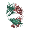
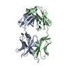
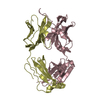
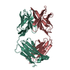
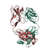
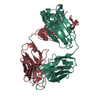
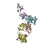
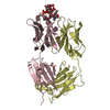
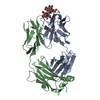
 Assembly

 Mass: 96.063 Da / Num. of mol.: 2 / Source method: obtained synthetically / Formula: SO4
 Mass: 18.015 Da / Num. of mol.: 4 / Source method: isolated from a natural source / Formula: H2O
 Sample preparation
 / Beamline: 08ID-1 / Wavelength: 0.9795
  Processing