 Movie
Movie Controller

[English] 日本語
 Yorodumi
- PDB-1rmf: STRUCTURES OF A MONOCLONAL ANTI-ICAM-1 ANTIBODY R6.5 FRAGMENT AT ... -
+ Open data

- Basic information
| Entry | Database: PDB / ID: 1rmf | ||||||
|---|---|---|---|---|---|---|---|
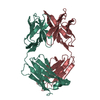
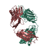
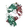
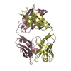
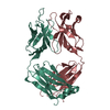
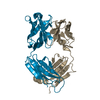
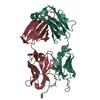
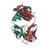
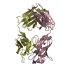
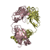
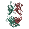
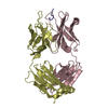
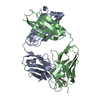
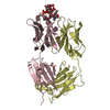
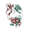
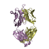
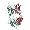
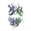
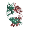
| Title | STRUCTURES OF A MONOCLONAL ANTI-ICAM-1 ANTIBODY R6.5 FRAGMENT AT 2.8 ANGSTROMS RESOLUTION | ||||||
 Components Components |
| ||||||
 Keywords Keywords | IMMUNOGLOBULIN | ||||||
| Function / homology | Immunoglobulins / Immunoglobulin-like / Sandwich / Mainly Beta / : / :  Function and homology information Function and homology information | ||||||
| Biological species |  | ||||||
| Method |  X-RAY DIFFRACTION / Resolution: 2.8 Å X-RAY DIFFRACTION / Resolution: 2.8 Å | ||||||
 Authors Authors | Jedrzejas, M.J. / Luo, M. | ||||||
 Citation Citation | Journal: Acta Crystallogr.,Sect.D / Year: 1995 Title: Structure of a monoclonal anti-ICAM-1 antibody R6.5 Fab fragment at 2.8 A resolution. Authors: Jedrzejas, M.J. / Miglietta, J. / Griffin, J.A. / Luo, M. | ||||||
| History |
|
- Structure visualization
| Structure viewer | Molecule: MolmilJmol/JSmol |
|---|
- Downloads & links
-Download
| PDBx/mmCIF format | 1rmf.cif.gz | 114 KB | Display | PDBx/mmCIF format |
|---|---|---|---|---|
| PDB format | pdb1rmf.ent.gz | 88.1 KB | Display | PDB format |
| PDBx/mmJSON format | 1rmf.json.gz | Tree view | PDBx/mmJSON format | |
| Others |  Other downloads Other downloads |
-Validation report
| Arichive directory | https://data.pdbj.org/pub/pdb/validation_reports/rm/1rmfftp://data.pdbj.org/pub/pdb/validation_reports/rm/1rmf | HTTPS FTP |
|---|
-Related structure data
| Similar structure data |
|---|

-Links
 PDBj
PDBj

- Assembly
| Deposited unit | 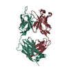
| ||||||||
|---|---|---|---|---|---|---|---|---|---|
| 1 |
| ||||||||
| Unit cell |
| ||||||||
| Atom site foot note | 1: CIS PROLINE - PRO L 8 / 2: CIS PROLINE - PRO L 100 / 3: CIS PROLINE - PRO L 146 / 4: CIS PROLINE - PRO H 9 5: ALA H 131 - PRO H 132 OMEGA = 220.96 PEPTIDE BOND DEVIATES SIGNIFICANTLY FROM TRANS CONFORMATION 6: CIS PROLINE - PRO H 153 / 7: CIS PROLINE - PRO H 155 8: TRP H 194 - PRO H 195 OMEGA = 51.63 PEPTIDE BOND DEVIATES SIGNIFICANTLY FROM TRANS CONFORMATION |
-Components
| #1: Antibody | Mass: 24068.684 Da / Num. of mol.: 1 Source method: isolated from a genetically manipulated source Source: (gene. exp.) |
|---|---|
| #2: Antibody | Mass: 23114.936 Da / Num. of mol.: 1 Source method: isolated from a genetically manipulated source Source: (gene. exp.) |
| #3: Water | ChemComp-HOH /  Mass: 18.015 Da / Num. of mol.: 39 / Source method: isolated from a natural source / Formula: H2O Mass: 18.015 Da / Num. of mol.: 39 / Source method: isolated from a natural source / Formula: H2O |
| Has protein modification | Y |
-Experimental details
-Experiment
| Experiment | Method: X-RAY DIFFRACTION |
|---|
- Sample preparation
| Crystal | Density Matthews: 2.69 Å3/Da / Density % sol: 54.26 % | |||||||||||||||
|---|---|---|---|---|---|---|---|---|---|---|---|---|---|---|---|---|
| Crystal grow | *PLUS pH: 8.5 / Method: other | |||||||||||||||
| Components of the solutions | *PLUS
|
-Data collection
| Reflection | Num. obs: 12598 / % possible obs: 84.1 % / Observed criterion σ(I): 2 |
|---|---|
| Reflection | *PLUS Observed criterion σ(I): 25.2 / Num. measured all: 32642 / Rmerge(I) obs: 0.064 |
- Processing
| Software |
| ||||||||||||||||||||||||||||||||||||||||||||||||||||||||||||
|---|---|---|---|---|---|---|---|---|---|---|---|---|---|---|---|---|---|---|---|---|---|---|---|---|---|---|---|---|---|---|---|---|---|---|---|---|---|---|---|---|---|---|---|---|---|---|---|---|---|---|---|---|---|---|---|---|---|---|---|---|---|
| Refinement | Resolution: 2.8→6.5 Å / σ(F): 4 /
| ||||||||||||||||||||||||||||||||||||||||||||||||||||||||||||
| Displacement parameters | Biso mean: 16.5 Å2 | ||||||||||||||||||||||||||||||||||||||||||||||||||||||||||||
| Refinement step | Cycle: LAST / Resolution: 2.8→6.5 Å
| ||||||||||||||||||||||||||||||||||||||||||||||||||||||||||||
| Refine LS restraints |
| ||||||||||||||||||||||||||||||||||||||||||||||||||||||||||||
| Refine LS restraints | *PLUS Type: x_dihedral_angle_deg / Dev ideal: 28.2 |
