 Movie
Movie Controller


+ Open data

- Basic information
| Entry | Database: PDB / ID: 1ae6 | ||||||
|---|---|---|---|---|---|---|---|
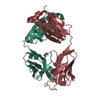
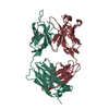
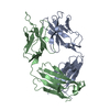
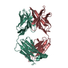
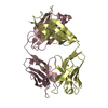
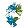
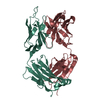
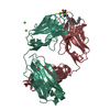
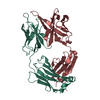
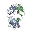
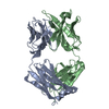
| Title | IGG-FAB FRAGMENT OF MOUSE MONOCLONAL ANTIBODY CTM01 | ||||||
 Components Components |
| ||||||
 Keywords Keywords | IMMUNOGLOBULIN / FAB FRAGMENT / HUMANISATION | ||||||
| Function / homology |  Function and homology information Function and homology informationInitial triggering of complement / Classical antibody-mediated complement activation / FCGR activation / Role of phospholipids in phagocytosis / Regulation of Complement cascade / phagocytosis, recognition / humoral immune response mediated by circulating immunoglobulin / Regulation of actin dynamics for phagocytic cup formation / positive regulation of type IIa hypersensitivity / positive regulation of type I hypersensitivity ...Initial triggering of complement / Classical antibody-mediated complement activation / FCGR activation / Role of phospholipids in phagocytosis / Regulation of Complement cascade / phagocytosis, recognition / humoral immune response mediated by circulating immunoglobulin / Regulation of actin dynamics for phagocytic cup formation / positive regulation of type IIa hypersensitivity / positive regulation of type I hypersensitivity / antibody-dependent cellular cytotoxicity / Fc-gamma receptor I complex binding / immunoglobulin complex, circulating / phagocytosis, engulfment / immunoglobulin receptor binding / IgG immunoglobulin complex / immunoglobulin mediated immune response / complement activation, classical pathway / immunoglobulin complex / antigen binding / positive regulation of phagocytosis / B cell differentiation / positive regulation of immune response / antibacterial humoral response / adaptive immune response / defense response to bacterium / external side of plasma membrane / : / metal ion binding / cytoplasm Similarity search - Function | ||||||
| Biological species |  | ||||||
| Method |  X-RAY DIFFRACTION / SYNCHROTRON / MOLECULAR REPLACEMENT / Resolution: 3 Å X-RAY DIFFRACTION / SYNCHROTRON / MOLECULAR REPLACEMENT / Resolution: 3 Å | ||||||
 Authors Authors | Banfield, M.J. / Brady, R.L. | ||||||
 Citation Citation | Journal: Proteins / Year: 1997 Title: VL:VH domain rotations in engineered antibodies: crystal structures of the Fab fragments from two murine antitumor antibodies and their engineered human constructs. Authors: Banfield, M.J. / King, D.J. / Mountain, A. / Brady, R.L. | ||||||
| History |
|
- Structure visualization
| Structure viewer | Molecule: MolmilJmol/JSmol |
|---|
- Downloads & links
-Download
| PDBx/mmCIF format | 1ae6.cif.gz | 88.9 KB | Display | PDBx/mmCIF format |
|---|---|---|---|---|
| PDB format | pdb1ae6.ent.gz | 67.4 KB | Display | PDB format |
| PDBx/mmJSON format | 1ae6.json.gz | Tree view | PDBx/mmJSON format | |
| Others |  Other downloads Other downloads |
-Validation report
| Arichive directory | https://data.pdbj.org/pub/pdb/validation_reports/ae/1ae6ftp://data.pdbj.org/pub/pdb/validation_reports/ae/1ae6 | HTTPS FTP |
|---|
-Related structure data
| Related structure data | 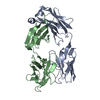 1ad0C 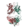 1ad9SC  1cloC S: Starting model for refinement C: citing same article ( |
|---|---|
| Similar structure data |


-Links
 PDBj
PDBj


- Assembly
| Deposited unit | 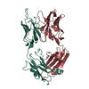
| ||||||||
|---|---|---|---|---|---|---|---|---|---|
| 1 |
| ||||||||
| Unit cell |
|
-Components
| #1: Antibody | Mass: 24194.924 Da / Num. of mol.: 1 / Fragment: FAB FRAGMENT / Source method: isolated from a natural source Details: EACH OF THE FOUR DOMAINS ADOPT THE IMMUNOGLOBULIN SUPERFAMILY FOLD Source: (natural) |
|---|---|
| #2: Antibody | Mass: 23469.332 Da / Num. of mol.: 1 / Fragment: FAB FRAGMENT / Source method: isolated from a natural source Details: EACH OF THE FOUR DOMAINS ADOPT THE IMMUNOGLOBULIN SUPERFAMILY FOLD Source: (natural) |
| Has protein modification | Y |
-Experimental details
-Experiment
| Experiment | Method: X-RAY DIFFRACTION / Number of used crystals: 5 |
|---|
- Sample preparation
| Crystal | Density Matthews: 2.26 Å3/Da / Density % sol: 37.5 % | |||||||||||||||||||||||||
|---|---|---|---|---|---|---|---|---|---|---|---|---|---|---|---|---|---|---|---|---|---|---|---|---|---|---|
| Crystal grow | pH: 5.6 Details: 20-25% PEG4000, BUFFERED AT PH 5.6 WITH 20MM BIS-TRIS. | |||||||||||||||||||||||||
| Crystal | *PLUS | |||||||||||||||||||||||||
| Crystal grow | *PLUS pH: 6 / Method: vapor diffusion, hanging drop | |||||||||||||||||||||||||
| Components of the solutions | *PLUS
|
-Data collection
| Diffraction | Mean temperature: 290 K |
|---|---|
| Diffraction source | Source: SYNCHROTRON / Site: SRS  / Beamline: PX7.2 / Wavelength: 1.488 / Beamline: PX7.2 / Wavelength: 1.488 |
| Detector | Type: MARRESEARCH / Detector: IMAGE PLATE / Date: Feb 1, 1995 / Details: MIRRORS |
| Radiation | Monochromator: MIRRORS / Monochromatic (M) / Laue (L): M / Scattering type: x-ray |
| Radiation wavelength | Wavelength: 1.488 Å / Relative weight: 1 |
| Reflection | Resolution: 3→20 Å / Num. obs: 7862 / % possible obs: 91.1 % / Observed criterion σ(I): 0 / Redundancy: 2 % / Biso Wilson estimate: 40.8 Å2 / Rsym value: 0.138 / Net I/σ(I): 5 |
| Reflection shell | Resolution: 3→3.23 Å / Mean I/σ(I) obs: 2.1 / Rsym value: 0.252 / % possible all: 88.3 |
| Reflection | *PLUS Num. measured all: 15707 / Rmerge(I) obs: 0.138 |
| Reflection shell | *PLUS % possible obs: 88.3 % / Rmerge(I) obs: 0.252 |
- Processing
| Software |
| ||||||||||||||||||||||||||||||||||||||||||||||||||||||||||||
|---|---|---|---|---|---|---|---|---|---|---|---|---|---|---|---|---|---|---|---|---|---|---|---|---|---|---|---|---|---|---|---|---|---|---|---|---|---|---|---|---|---|---|---|---|---|---|---|---|---|---|---|---|---|---|---|---|---|---|---|---|---|
| Refinement | Method to determine structure: MOLECULAR REPLACEMENT Starting model: PDB ENTRY 1AD9 - STRUCTURE OF THE ENGINEERED HUMAN CONSTRUCT OF CTM01 Resolution: 3→15 Å / Rfactor Rfree error: 0.013 / Data cutoff high absF: 10000000 / Data cutoff low absF: 0.001 / Isotropic thermal model: GROUP / Cross valid method: THROUGHOUT / σ(F): 0 Details: BULK SOLVENT MODEL USED ELECTRON DENSITY IS WEAK FOR RESIDUES H 96 - H 100A OF THE H3 HYPERVARIABLE LOOP. ALL ATOMS FOR THIS LOOP ARE INCLUDED IN THE FINAL MODEL, BUT THE DENSITY ONLY ...Details: BULK SOLVENT MODEL USED ELECTRON DENSITY IS WEAK FOR RESIDUES H 96 - H 100A OF THE H3 HYPERVARIABLE LOOP. ALL ATOMS FOR THIS LOOP ARE INCLUDED IN THE FINAL MODEL, BUT THE DENSITY ONLY ACCOUNTS FOR THE C-ALPHA TRACE.
| ||||||||||||||||||||||||||||||||||||||||||||||||||||||||||||
| Displacement parameters | Biso mean: 44.6 Å2 | ||||||||||||||||||||||||||||||||||||||||||||||||||||||||||||
| Refine analyze |
| ||||||||||||||||||||||||||||||||||||||||||||||||||||||||||||
| Refinement step | Cycle: LAST / Resolution: 3→15 Å
| ||||||||||||||||||||||||||||||||||||||||||||||||||||||||||||
| Refine LS restraints |
| ||||||||||||||||||||||||||||||||||||||||||||||||||||||||||||
| LS refinement shell | Resolution: 3→3.14 Å / Rfactor Rfree error: 0.06 / Total num. of bins used: 8
| ||||||||||||||||||||||||||||||||||||||||||||||||||||||||||||
| Xplor file |
| ||||||||||||||||||||||||||||||||||||||||||||||||||||||||||||
| Software | *PLUS Name: X-PLOR / Version: 3.851 / Classification: refinement | ||||||||||||||||||||||||||||||||||||||||||||||||||||||||||||
| Refinement | *PLUS Rfactor obs: 0.21 / Rfactor Rwork: 0.21 | ||||||||||||||||||||||||||||||||||||||||||||||||||||||||||||
| Solvent computation | *PLUS | ||||||||||||||||||||||||||||||||||||||||||||||||||||||||||||
| Displacement parameters | *PLUS | ||||||||||||||||||||||||||||||||||||||||||||||||||||||||||||
| Refine LS restraints | *PLUS
|
