 Movie
Movie Controller

[English] 日本語
 Yorodumi
- PDB-4mhj: Crystal structure of Fab H5M9 in complex with influenza virus hem... -
+ Open data

- Basic information
| Entry | Database: PDB / ID: 4mhj | |||||||||
|---|---|---|---|---|---|---|---|---|---|---|
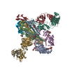
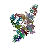
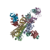
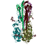
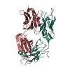
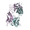
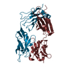
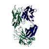
| Title | Crystal structure of Fab H5M9 in complex with influenza virus hemagglutinin from A/goose/Guangdong/1/96 (H5N1) | |||||||||
 Components Components |
| |||||||||
 Keywords Keywords | VIRAL PROTEIN/IMMUNE SYSTEM / H5N1 influenza virus / epitope / broadly neutralizing antibody / VIRAL PROTEIN-IMMUNE SYSTEM complex | |||||||||
| Function / homology |  Function and homology information Function and homology informationviral budding from plasma membrane / clathrin-dependent endocytosis of virus by host cell / host cell surface receptor binding / fusion of virus membrane with host plasma membrane / fusion of virus membrane with host endosome membrane / viral envelope / virion attachment to host cell / host cell plasma membrane / virion membrane / membrane Similarity search - Function | |||||||||
| Biological species |   Influenza A virus Influenza A virus | |||||||||
| Method |  X-RAY DIFFRACTION / SYNCHROTRON / MOLECULAR REPLACEMENT / Resolution: 6.98 Å X-RAY DIFFRACTION / SYNCHROTRON / MOLECULAR REPLACEMENT / Resolution: 6.98 Å | |||||||||
 Authors Authors | Zhu, X. / Wilson, I.A. | |||||||||
 Citation Citation | Journal: J.Virol. / Year: 2013 Title: A Unique and Conserved Neutralization Epitope in H5N1 Influenza Viruses Identified by an Antibody against the A/Goose/Guangdong/1/96 Hemagglutinin. Authors: Zhu, X. / Guo, Y.H. / Jiang, T. / Wang, Y.D. / Chan, K.H. / Li, X.F. / Yu, W. / McBride, R. / Paulson, J.C. / Yuen, K.Y. / Qin, C.F. / Che, X.Y. / Wilson, I.A. | |||||||||
| History |
|
- Structure visualization
| Structure viewer | Molecule: MolmilJmol/JSmol |
|---|
- Downloads & links
-Download
| PDBx/mmCIF format | 4mhj.cif.gz | 938.4 KB | Display | PDBx/mmCIF format |
|---|---|---|---|---|
| PDB format | pdb4mhj.ent.gz | 753.3 KB | Display | PDB format |
| PDBx/mmJSON format | 4mhj.json.gz | Tree view | PDBx/mmJSON format | |
| Others |  Other downloads Other downloads |
-Validation report
| Arichive directory | https://data.pdbj.org/pub/pdb/validation_reports/mh/4mhjftp://data.pdbj.org/pub/pdb/validation_reports/mh/4mhj | HTTPS FTP |
|---|
-Related structure data
| Related structure data |  4mhhC  4mhiSC  3gbmS C: citing same article ( S: Starting model for refinement |
|---|---|
| Similar structure data |
-Links
 PDBj
PDBj


- Assembly


-Components
-Hemagglutinin ... , 2 types, 12 molecules ACGMOSBDNPIU
| #1: Protein | Mass: 37812.707 Da / Num. of mol.: 6 / Fragment: receptor binding domain (UNP residues 17-346) Source method: isolated from a genetically manipulated source Source: (gene. exp.) Influenza A virus / Strain: A/Goose/Guangdong/1/1996 H5N1 genotype Gs/Gd / Gene: HA / Plasmid: pfastbac-HT / Production host:  Trichoplusia ni (cabbage looper) / Strain (production host): Hi5 / References: UniProt: Q9Q0U6 Trichoplusia ni (cabbage looper) / Strain (production host): Hi5 / References: UniProt: Q9Q0U6#2: Protein | Mass: 20894.012 Da / Num. of mol.: 6 / Fragment: membrane fusion domain (UNP residues 347-521) Source method: isolated from a genetically manipulated source Source: (gene. exp.) Influenza A virus / Strain: A/Goose/Guangdong/1/1996 H5N1 genotype Gs/Gd / Gene: HA / Plasmid: pfastbac-HT / Production host: Trichoplusia ni (cabbage looper) / Strain (production host): Hi5 / References: UniProt: Q9Q0U6 |
|---|
-Antibody , 2 types, 12 molecules LEJXQVHFKTRW
| #3: Antibody | Mass: 24049.486 Da / Num. of mol.: 6 / Fragment: Fab Source method: isolated from a genetically manipulated source Source: (gene. exp.) #4: Antibody | Mass: 23935.797 Da / Num. of mol.: 6 / Fragment: Fab Source method: isolated from a genetically manipulated source Source: (gene. exp.) |
|---|
-Sugars , 6 types, 14 molecules 
| #5: Polysaccharide | Source method: isolated from a genetically manipulated source #6: Polysaccharide | alpha-D-mannopyranose-(1-2)-alpha-D-mannopyranose-(1-3)-[alpha-D-mannopyranose-(1-2)-alpha-D- ...alpha-D-mannopyranose-(1-2)-alpha-D-mannopyranose-(1-3)-[alpha-D-mannopyranose-(1-2)-alpha-D-mannopyranose-(1-6)]alpha-D-mannopyranose-(1-6)-beta-D-mannopyranose-(1-4)-2-acetamido-2-deoxy-beta-D-glucopyranose-(1-4)-2-acetamido-2-deoxy-beta-D-glucopyranose | Source method: isolated from a genetically manipulated source #7: Polysaccharide | Source method: isolated from a genetically manipulated source #8: Polysaccharide | beta-D-mannopyranose-(1-4)-2-acetamido-2-deoxy-beta-D-glucopyranose-(1-4)-2-acetamido-2-deoxy-beta- ...beta-D-mannopyranose-(1-4)-2-acetamido-2-deoxy-beta-D-glucopyranose-(1-4)-2-acetamido-2-deoxy-beta-D-glucopyranose | Source method: isolated from a genetically manipulated source #9: Polysaccharide | alpha-D-mannopyranose-(1-3)-beta-D-mannopyranose-(1-4)-2-acetamido-2-deoxy-beta-D-glucopyranose-(1- ...alpha-D-mannopyranose-(1-3)-beta-D-mannopyranose-(1-4)-2-acetamido-2-deoxy-beta-D-glucopyranose-(1-4)-2-acetamido-2-deoxy-beta-D-glucopyranose | Source method: isolated from a genetically manipulated source #10: Sugar | ChemComp-NAG /  Type: D-saccharide, beta linking / Mass: 221.208 Da / Num. of mol.: 6 Type: D-saccharide, beta linking / Mass: 221.208 Da / Num. of mol.: 6Source method: isolated from a genetically manipulated source Formula: C8H15NO6 |
|---|
-Details
| Has protein modification | Y |
|---|
-Experimental details
-Experiment
| Experiment | Method: X-RAY DIFFRACTION / Number of used crystals: 1 |
|---|
- Sample preparation
| Crystal | Density Matthews: 4.19 Å3/Da / Density % sol: 70.66 % |
|---|---|
| Crystal grow | Temperature: 295 K / Method: vapor diffusion, sitting drop / pH: 7.5 Details: 0.1 M HEPES, pH 7.5, 2% PEG400, 2.0 M ammonium sulfate, VAPOR DIFFUSION, SITTING DROP, temperature 295K |
-Data collection
| Diffraction | Mean temperature: 100 K |
|---|---|
| Diffraction source | Source: SYNCHROTRON / Site: SSRL  / Beamline: BL11-1 / Wavelength: 0.97945 Å / Beamline: BL11-1 / Wavelength: 0.97945 Å |
| Detector | Type: MARMOSAIC 325 mm CCD / Detector: CCD / Date: Mar 19, 2010 |
| Radiation | Monochromator: Side scattering bent cube-root I-beam single crystal, asymmetric cut 4.965 degrees Protocol: SINGLE WAVELENGTH / Monochromatic (M) / Laue (L): M / Scattering type: x-ray |
| Radiation wavelength | Wavelength: 0.97945 Å / Relative weight: 1 |
| Reflection | Resolution: 6.979→174.078 Å / Num. obs: 16538 / % possible obs: 93.3 % / Observed criterion σ(I): -3 / Redundancy: 5.8 % / Rsym value: 0.14 / Net I/σ(I): 12.9 |
| Reflection shell | Resolution: 6.979→7.44 Å / Redundancy: 6.2 % / Mean I/σ(I) obs: 1.2 / Rsym value: 0.92 / % possible all: 64 |
- Processing
| Software |
| ||||||||||||||||||||
|---|---|---|---|---|---|---|---|---|---|---|---|---|---|---|---|---|---|---|---|---|---|
| Refinement | Method to determine structure: MOLECULAR REPLACEMENT Starting model: PDB ENTRIES 3GBM AND 4MHI Resolution: 6.98→50.08 Å / Cor.coef. Fo:Fc: 0.759 / Cor.coef. Fo:Fc free: 0.792 / SU B: 0.008 / SU ML: 0 / Cross valid method: THROUGHOUT / ESU R: 4.762 / ESU R Free: 4.646 / Stereochemistry target values: MAXIMUM LIKELIHOOD / Details: HYDROGENS HAVE BEEN ADDED IN THE RIDING POSITIONS
| ||||||||||||||||||||
| Solvent computation | Ion probe radii: 0.8 Å / Shrinkage radii: 0.8 Å / VDW probe radii: 1.2 Å / Solvent model: MASK | ||||||||||||||||||||
| Displacement parameters | Biso mean: 89.86 Å2
| ||||||||||||||||||||
| Refinement step | Cycle: LAST / Resolution: 6.98→50.08 Å
| ||||||||||||||||||||
| LS refinement shell | Resolution: 6.98→7.16 Å / Total num. of bins used: 20
|
