 Movie
Movie Controller

[English] 日本語
 Yorodumi
- PDB-3whe: A new conserved neutralizing epitope at the globular head of hema... -
+ Open data

- Basic information
| Entry | Database: PDB / ID: 3whe | |||||||||
|---|---|---|---|---|---|---|---|---|---|---|
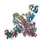
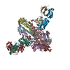
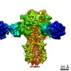
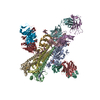
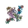
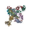
| Title | A new conserved neutralizing epitope at the globular head of hemagglutinin in H3N2 influenza viruses | |||||||||
 Components Components |
| |||||||||
 Keywords Keywords | VIRAL PROTEIN/IMMUNE SYSTEM / VIRAL PROTEIN-IMMUNE SYSTEM complex | |||||||||
| Function / homology |  Function and homology information Function and homology informationviral budding from plasma membrane / clathrin-dependent endocytosis of virus by host cell / host cell surface receptor binding / fusion of virus membrane with host plasma membrane / fusion of virus membrane with host endosome membrane / viral envelope / virion attachment to host cell / host cell plasma membrane / virion membrane / membrane Similarity search - Function | |||||||||
| Biological species |   Influenza A virus Influenza A virus Homo sapiens (human) Homo sapiens (human) | |||||||||
| Method |  X-RAY DIFFRACTION / SYNCHROTRON / MOLECULAR REPLACEMENT / Resolution: 4 Å X-RAY DIFFRACTION / SYNCHROTRON / MOLECULAR REPLACEMENT / Resolution: 4 Å | |||||||||
 Authors Authors | Fujii, Y. / Sumida, T. / Shirouzu, M. / Yokoyama, S. | |||||||||
 Citation Citation | Journal: J.Virol. / Year: 2014 Title: Conserved neutralizing epitope at globular head of hemagglutinin in H3N2 influenza viruses. Authors: Iba, Y. / Fujii, Y. / Ohshima, N. / Sumida, T. / Kubota-Koketsu, R. / Ikeda, M. / Wakiyama, M. / Shirouzu, M. / Okada, J. / Okuno, Y. / Kurosawa, Y. / Yokoyama, S. | |||||||||
| History |
|
- Structure visualization
| Structure viewer | Molecule: MolmilJmol/JSmol |
|---|
- Downloads & links
-Download
| PDBx/mmCIF format | 3whe.cif.gz | 1.9 MB | Display | PDBx/mmCIF format |
|---|---|---|---|---|
| PDB format | pdb3whe.ent.gz | Display | PDB format | |
| PDBx/mmJSON format | 3whe.json.gz | Tree view | PDBx/mmJSON format | |
| Others |  Other downloads Other downloads |
-Validation report
| Arichive directory | https://data.pdbj.org/pub/pdb/validation_reports/wh/3wheftp://data.pdbj.org/pub/pdb/validation_reports/wh/3whe | HTTPS FTP |
|---|
-Related structure data
| Related structure data | 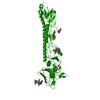 1ha0S S: Starting model for refinement |
|---|---|
| Similar structure data |


-Links
 PDBj
PDBj


- Assembly
| Deposited unit | 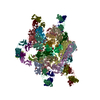
| ||||||||
|---|---|---|---|---|---|---|---|---|---|
| 1 | 
| ||||||||
| 2 | 
| ||||||||
| 3 | 
| ||||||||
| 4 | 
| ||||||||
| Unit cell |
|
-Components
-Protein , 1 types, 12 molecules ABCDEFGHIJKL
| #1: Protein | Mass: 55221.602 Da / Num. of mol.: 12 / Fragment: UNP RESIDUES 25-517 Source method: isolated from a genetically manipulated source Source: (gene. exp.) Influenza A virus / Strain: A/Aichi/2-1/1968 (H3N2) / Gene: 126567434, HA / Plasmid: pBAC-3 / Cell line (production host): SF+ / Production host:   Spodoptera frugiperda (fall armyworm) / References: UniProt: T2HNI1 Spodoptera frugiperda (fall armyworm) / References: UniProt: T2HNI1 |
|---|
-Antibody , 2 types, 24 molecules MOQSUWY13579NPRTVXZ24680
| #2: Antibody | Mass: 24187.277 Da / Num. of mol.: 12 Source method: isolated from a genetically manipulated source Source: (gene. exp.) Homo sapiens (human) / Description: protein selected by phage display / Plasmid: pCMVscript / Production host:   Cricetulus griseus (Chinese hamster) / Strain (production host): CHO-K1 Cricetulus griseus (Chinese hamster) / Strain (production host): CHO-K1#3: Antibody | Mass: 22938.275 Da / Num. of mol.: 12 Source method: isolated from a genetically manipulated source Source: (gene. exp.) Homo sapiens (human) / Description: protein selected by phage display / Plasmid: pCMVscript / Production host: Cricetulus griseus (Chinese hamster) / Strain (production host): CHO-K1 |
|---|
-Sugars , 4 types, 60 molecules 
| #4: Polysaccharide | alpha-D-mannopyranose-(1-3)-[alpha-D-mannopyranose-(1-6)]alpha-D-mannopyranose-(1-6)-[alpha-D- ...alpha-D-mannopyranose-(1-3)-[alpha-D-mannopyranose-(1-6)]alpha-D-mannopyranose-(1-6)-[alpha-D-mannopyranose-(1-3)]beta-D-mannopyranose-(1-4)-2-acetamido-2-deoxy-beta-D-glucopyranose-(1-4)-2-acetamido-2-deoxy-beta-D-glucopyranose Source method: isolated from a genetically manipulated source #5: Polysaccharide | alpha-D-mannopyranose-(1-2)-alpha-D-mannopyranose-(1-3)-[alpha-D-mannopyranose-(1-6)]beta-D- ...alpha-D-mannopyranose-(1-2)-alpha-D-mannopyranose-(1-3)-[alpha-D-mannopyranose-(1-6)]beta-D-mannopyranose-(1-4)-2-acetamido-2-deoxy-beta-D-glucopyranose-(1-4)-2-acetamido-2-deoxy-beta-D-glucopyranose Source method: isolated from a genetically manipulated source #6: Polysaccharide | beta-D-mannopyranose-(1-4)-2-acetamido-2-deoxy-beta-D-glucopyranose-(1-4)-2-acetamido-2-deoxy-beta- ...beta-D-mannopyranose-(1-4)-2-acetamido-2-deoxy-beta-D-glucopyranose-(1-4)-2-acetamido-2-deoxy-beta-D-glucopyranose Source method: isolated from a genetically manipulated source #7: Sugar | ChemComp-NAG /  Type: D-saccharide, beta linking / Mass: 221.208 Da / Num. of mol.: 24 Type: D-saccharide, beta linking / Mass: 221.208 Da / Num. of mol.: 24Source method: isolated from a genetically manipulated source Formula: C8H15NO6 |
|---|
-Details
| Has protein modification | Y |
|---|
-Experimental details
-Experiment
| Experiment | Method: X-RAY DIFFRACTION / Number of used crystals: 1 |
|---|
- Sample preparation
| Crystal | Density Matthews: 3.57 Å3/Da / Density % sol: 65.53 % |
|---|---|
| Crystal grow | Temperature: 293 K / Method: vapor diffusion, sitting drop / pH: 5.5 Details: PEG8000, KCl, pH 5.5, VAPOR DIFFUSION, SITTING DROP, temperature 293K |
-Data collection
| Diffraction | Mean temperature: 100 K | ||||||||||||||||||||||||||||||||||||||||||||||||||||||||||||||||||||||||||||||||||||||||
|---|---|---|---|---|---|---|---|---|---|---|---|---|---|---|---|---|---|---|---|---|---|---|---|---|---|---|---|---|---|---|---|---|---|---|---|---|---|---|---|---|---|---|---|---|---|---|---|---|---|---|---|---|---|---|---|---|---|---|---|---|---|---|---|---|---|---|---|---|---|---|---|---|---|---|---|---|---|---|---|---|---|---|---|---|---|---|---|---|---|
| Diffraction source | Source: SYNCHROTRON / Site: SPring-8  / Beamline: BL41XU / Wavelength: 1 Å / Beamline: BL41XU / Wavelength: 1 Å | ||||||||||||||||||||||||||||||||||||||||||||||||||||||||||||||||||||||||||||||||||||||||
| Detector | Type: RAYONIX MX225HE / Detector: CCD / Date: Oct 22, 2011 | ||||||||||||||||||||||||||||||||||||||||||||||||||||||||||||||||||||||||||||||||||||||||
| Radiation | Monochromator: Rotated-inclined double-crystal monochromator Protocol: SINGLE WAVELENGTH / Monochromatic (M) / Laue (L): M / Scattering type: x-ray | ||||||||||||||||||||||||||||||||||||||||||||||||||||||||||||||||||||||||||||||||||||||||
| Radiation wavelength | Wavelength: 1 Å / Relative weight: 1 | ||||||||||||||||||||||||||||||||||||||||||||||||||||||||||||||||||||||||||||||||||||||||
| Reflection | Resolution: 4→30 Å / Num. obs: 142111 / % possible obs: 98 % / Observed criterion σ(F): 0 / Observed criterion σ(I): -3 / Redundancy: 3.905 % / Rmerge(I) obs: 0.236 / Net I/σ(I): 6.35 | ||||||||||||||||||||||||||||||||||||||||||||||||||||||||||||||||||||||||||||||||||||||||
| Reflection shell | Rmerge(I) obs: 2.1 / Diffraction-ID: 1
|
- Processing
| Software |
| |||||||||||||||||||||||||||||||||||||||||||||||||||||||||||||||||||||||||||||||||||||||||||||||||||
|---|---|---|---|---|---|---|---|---|---|---|---|---|---|---|---|---|---|---|---|---|---|---|---|---|---|---|---|---|---|---|---|---|---|---|---|---|---|---|---|---|---|---|---|---|---|---|---|---|---|---|---|---|---|---|---|---|---|---|---|---|---|---|---|---|---|---|---|---|---|---|---|---|---|---|---|---|---|---|---|---|---|---|---|---|---|---|---|---|---|---|---|---|---|---|---|---|---|---|---|---|
| Refinement | Method to determine structure: MOLECULAR REPLACEMENT Starting model: 1HA0 Resolution: 4→30 Å / Rfactor Rfree error: 0.004 / Occupancy max: 1 / Occupancy min: 1 / Data cutoff high absF: 17247948 / Data cutoff low absF: 0 / Isotropic thermal model: RESTRAINED / Cross valid method: THROUGHOUT / σ(F): 0 / Stereochemistry target values: Engh & Huber / Details: BULK SOLVENT MODEL USED
| |||||||||||||||||||||||||||||||||||||||||||||||||||||||||||||||||||||||||||||||||||||||||||||||||||
| Solvent computation | Solvent model: FLAT MODEL / Bsol: 146.958 Å2 / ksol: 0.3 e/Å3 | |||||||||||||||||||||||||||||||||||||||||||||||||||||||||||||||||||||||||||||||||||||||||||||||||||
| Displacement parameters | Biso max: 381.87 Å2 / Biso mean: 205.8505 Å2 / Biso min: 87.44 Å2
| |||||||||||||||||||||||||||||||||||||||||||||||||||||||||||||||||||||||||||||||||||||||||||||||||||
| Refine analyze |
| |||||||||||||||||||||||||||||||||||||||||||||||||||||||||||||||||||||||||||||||||||||||||||||||||||
| Refinement step | Cycle: LAST / Resolution: 4→30 Å
| |||||||||||||||||||||||||||||||||||||||||||||||||||||||||||||||||||||||||||||||||||||||||||||||||||
| Refine LS restraints |
| |||||||||||||||||||||||||||||||||||||||||||||||||||||||||||||||||||||||||||||||||||||||||||||||||||
| LS refinement shell | Refine-ID: X-RAY DIFFRACTION / Total num. of bins used: 10
| |||||||||||||||||||||||||||||||||||||||||||||||||||||||||||||||||||||||||||||||||||||||||||||||||||
| Xplor file |
|
