 Movie
Movie Controller


+ Open data

- Basic information
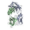
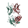
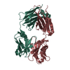
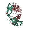
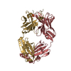
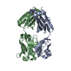
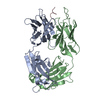
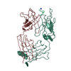
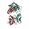
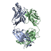
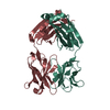
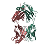
| Entry | Database: PDB / ID: 1clo | ||||||
|---|---|---|---|---|---|---|---|
| Title | ANTI-CARCINOEMBRYONIC ANTIGEN MONOCLONAL ANTIBODY A5B7 | ||||||
 Components Components | (A5B7 MONOCLONAL ANTIBODY) x 2 | ||||||
 Keywords Keywords | IMMUNOGLOBULIN / FAB-FRAGMENT | ||||||
| Function / homology |  Function and homology information Function and homology information | ||||||
| Biological species |  | ||||||
| Method |  X-RAY DIFFRACTION / SYNCHROTRON / MOLECULAR REPLACEMENT / Resolution: 2.1 Å X-RAY DIFFRACTION / SYNCHROTRON / MOLECULAR REPLACEMENT / Resolution: 2.1 Å | ||||||
 Authors Authors | Banfield, M.J. / King, D.J. / Mountain, A. / Brady, R.L. | ||||||
 Citation Citation | Journal: Proteins / Year: 1997 Title: VL:VH domain rotations in engineered antibodies: crystal structures of the Fab fragments from two murine antitumor antibodies and their engineered human constructs. Authors: Banfield, M.J. / King, D.J. / Mountain, A. / Brady, R.L. #1: Journal: To be PublishedTitle: VL:VH Domain Rotations in Engineeded Antibodies: Crystal Structures of the Fab Fragments from Two Murine Anti-Tumour Antibodies and Their Engineered Human Constructs Authors: Banfield, M.J. / King, D.J. / Mountain, A. / Brady, R.L. | ||||||
| History |
|
- Structure visualization
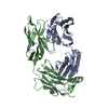
| Structure viewer | Molecule: MolmilJmol/JSmol |
|---|
- Downloads & links
-Download
| PDBx/mmCIF format | 1clo.cif.gz | 105.9 KB | Display | PDBx/mmCIF format |
|---|---|---|---|---|
| PDB format | pdb1clo.ent.gz | 79.1 KB | Display | PDB format |
| PDBx/mmJSON format | 1clo.json.gz | Tree view | PDBx/mmJSON format | |
| Others |  Other downloads Other downloads |
-Validation report
| Arichive directory | https://data.pdbj.org/pub/pdb/validation_reports/cl/1cloftp://data.pdbj.org/pub/pdb/validation_reports/cl/1clo | HTTPS FTP |
|---|
-Related structure data


-Links
 PDBj
PDBj

- Assembly
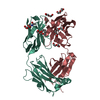
| Deposited unit | 
| ||||||||
|---|---|---|---|---|---|---|---|---|---|
| 1 |
| ||||||||
| Unit cell |
|
-Components
| #1: Antibody | Mass: 23238.602 Da / Num. of mol.: 1 / Fragment: ANTIGEN BINDING FRAGMENT, FAB / Source method: isolated from a natural source Details: ANTI-CARCINOEMBRYONIC ANTIGEN MONOCLONAL ANTIBODY A5B7 Source: (natural) |
|---|---|
| #2: Antibody | Mass: 24067.021 Da / Num. of mol.: 1 / Fragment: ANTIGEN BINDING FRAGMENT, FAB / Source method: isolated from a natural source Details: ANTI-CARCINOEMBRYONIC ANTIGEN MONOCLONAL ANTIBODY A5B7 Source: (natural) |
| #3: Water | ChemComp-HOH /  Mass: 18.015 Da / Num. of mol.: 400 / Source method: isolated from a natural source / Formula: H2O Mass: 18.015 Da / Num. of mol.: 400 / Source method: isolated from a natural source / Formula: H2O |
| Has protein modification | Y |
| Sequence details | THE FAB FRAGMENT IS NUMBERED ACCORDING TO THE KABAT SYSTEM (E.A.KABAT, T.T.WU,M.REID-MILLER,H.M. ...THE FAB FRAGMENT IS NUMBERED ACCORDING TO THE KABAT SYSTEM (E.A.KABAT, T.T.WU,M.REID-MILLER,H.M.PERRY, K.S.GOTTESMAN (1987) SEQUENCES OF PROTEINS OF IMMUNOLOGI |
-Experimental details
-Experiment
| Experiment | Method: X-RAY DIFFRACTION / Number of used crystals: 1 |
|---|
- Sample preparation
| Crystal | Density Matthews: 2.17 Å3/Da / Density % sol: 43.25 % | |||||||||||||||||||||||||
|---|---|---|---|---|---|---|---|---|---|---|---|---|---|---|---|---|---|---|---|---|---|---|---|---|---|---|
| Crystal grow | *PLUS pH: 6 / Method: vapor diffusion, hanging drop / Details: Banfield, M.J., (1996) Acta Cryst. D., 52, 1107. | |||||||||||||||||||||||||
| Components of the solutions | *PLUS
|
-Data collection
| Diffraction | Mean temperature: 100 K |
|---|---|
| Diffraction source | Source: SYNCHROTRON / Site: SRS  / Beamline: PX7.2 / Wavelength: 1.488 / Beamline: PX7.2 / Wavelength: 1.488 |
| Detector | Type: MAR scanner 180 mm plate / Detector: IMAGE PLATE / Date: Jun 20, 1995 |
| Radiation | Monochromatic (M) / Laue (L): M / Scattering type: x-ray |
| Radiation wavelength | Wavelength: 1.488 Å / Relative weight: 1 |
| Reflection | Resolution: 2→20 Å / Num. obs: 27439 / % possible obs: 99.2 % / Observed criterion σ(I): -3 / Redundancy: 3.5 % / Rmerge(I) obs: 0.047 / Net I/σ(I): 24.1 |
| Reflection shell | Resolution: 2→2.15 Å / Rmerge(I) obs: 0.115 / Mean I/σ(I) obs: 9.7 / % possible all: 98 |
| Reflection | *PLUS Num. measured all: 95102 |
| Reflection shell | *PLUS % possible obs: 98 % |
- Processing
| Software |
| ||||||||||||||||||||||||||||||||||||||||||||||||||||||||||||
|---|---|---|---|---|---|---|---|---|---|---|---|---|---|---|---|---|---|---|---|---|---|---|---|---|---|---|---|---|---|---|---|---|---|---|---|---|---|---|---|---|---|---|---|---|---|---|---|---|---|---|---|---|---|---|---|---|---|---|---|---|---|
| Refinement | Method to determine structure: MOLECULAR REPLACEMENT / Resolution: 2.1→8 Å / σ(F): 0 Details: WEAK ELECTRON DENSITY ONLY IS PRESENT FOR RESIDUES L 155 - L 159 AND H 130 - H 134.
| ||||||||||||||||||||||||||||||||||||||||||||||||||||||||||||
| Displacement parameters | Biso mean: 23.67 Å2 | ||||||||||||||||||||||||||||||||||||||||||||||||||||||||||||
| Refinement step | Cycle: LAST / Resolution: 2.1→8 Å
| ||||||||||||||||||||||||||||||||||||||||||||||||||||||||||||
| Refine LS restraints |
|
