 Movie
Movie Controller


+ Open data

- Basic information
| Entry | Database: PDB / ID: 1e6o | ||||||
|---|---|---|---|---|---|---|---|
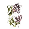
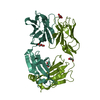
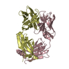
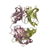
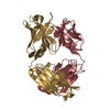
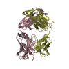
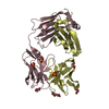
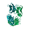
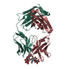
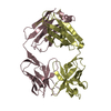
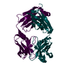
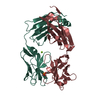
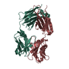
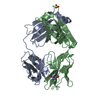
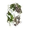
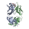
| Title | Crystal structure of Fab13B5 against HIV-1 capsid protein p24 | ||||||
 Components Components |
| ||||||
 Keywords Keywords | IMMUNOGLOBULIN / FAB / ANTIBODY / ANTIGEN / HIV-1 / P24 / CA | ||||||
| Function / homology | Immunoglobulins / Immunoglobulin-like / Sandwich / Mainly Beta Function and homology information Function and homology information | ||||||
| Biological species |  | ||||||
| Method |  X-RAY DIFFRACTION / SYNCHROTRON / MOLECULAR REPLACEMENT / Resolution: 1.8 Å X-RAY DIFFRACTION / SYNCHROTRON / MOLECULAR REPLACEMENT / Resolution: 1.8 Å | ||||||
 Authors Authors | Monaco-Malbet, S. / Berthet-Colominas, C. / Novelli, A. / Battai, N. / Piga, N. / Mallet, F. / Cusack, S. | ||||||
 Citation Citation | Journal: Structure / Year: 2000 Title: Mutual Conformational Adaptations in Antigen and Antibody Upon Complex Formation between an Fab and HIV-1 Capsid Protein P24 Authors: Monaco-Malbet, S. / Berthet-Colominas, C. / Novelli, A. / Battai, N. / Piga, N. / Cheynet, V. / Mallet, F. / Cusack, S. | ||||||
| History |
|
- Structure visualization
| Structure viewer | Molecule: MolmilJmol/JSmol |
|---|
- Downloads & links
-Download
| PDBx/mmCIF format | 1e6o.cif.gz | 98.3 KB | Display | PDBx/mmCIF format |
|---|---|---|---|---|
| PDB format | pdb1e6o.ent.gz | 74.5 KB | Display | PDB format |
| PDBx/mmJSON format | 1e6o.json.gz | Tree view | PDBx/mmJSON format | |
| Others |  Other downloads Other downloads |
-Validation report
| Arichive directory | https://data.pdbj.org/pub/pdb/validation_reports/e6/1e6oftp://data.pdbj.org/pub/pdb/validation_reports/e6/1e6o | HTTPS FTP |
|---|
-Related structure data
| Related structure data |  1e6jC  1afvS C: citing same article ( S: Starting model for refinement |
|---|---|
| Similar structure data |


-Links
 PDBj
PDBj


- Assembly
| Deposited unit | 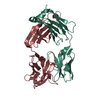
| ||||||||
|---|---|---|---|---|---|---|---|---|---|
| 1 |
| ||||||||
| Unit cell |
|
-Components
| #1: Antibody | Mass: 23552.301 Da / Num. of mol.: 1 / Fragment: HEAVY CHAIN 1-219 / Source method: isolated from a natural source / Details: OBTAINED BY PEPSIN CLEAVAGE (FAB') / Source: (natural) |
|---|---|
| #2: Antibody | Mass: 23285.732 Da / Num. of mol.: 1 / Fragment: LIGHT CHAIN 1-210 / Source method: isolated from a natural source / Details: OBTAINED BY PEPSIN CLEAVAGE (FAB') / Source: (natural) |
| #3: Water | ChemComp-HOH /  Mass: 18.015 Da / Num. of mol.: 237 / Source method: isolated from a natural source / Formula: H2O Mass: 18.015 Da / Num. of mol.: 237 / Source method: isolated from a natural source / Formula: H2O |
| Has protein modification | Y |
-Experimental details
-Experiment
| Experiment | Method: X-RAY DIFFRACTION / Number of used crystals: 1 |
|---|
- Sample preparation
| Crystal | Density Matthews: 2.2 Å3/Da / Density % sol: 44 % | |||||||||||||||
|---|---|---|---|---|---|---|---|---|---|---|---|---|---|---|---|---|
| Crystal grow | Temperature: 295 K / Method: vapor diffusion, hanging drop / pH: 7.8 Details: 7MG/ML OF FAB'13B5 IN 0.1M PIPES PH=7.8 IN 9% TO 11% PEG8000 AFTER EQUILIBRATION IN HANGING DROPS AT 22C, pH 7.80 | |||||||||||||||
| Crystal grow | *PLUS Temperature: 22 ℃ / Method: vapor diffusion, hanging drop | |||||||||||||||
| Components of the solutions | *PLUS
|
-Data collection
| Diffraction | Mean temperature: 100 K |
|---|---|
| Diffraction source | Source: SYNCHROTRON / Site: ESRF  / Beamline: BM14 / Wavelength: 0.799 / Beamline: BM14 / Wavelength: 0.799 |
| Detector | Type: MARRESEARCH / Detector: IMAGE PLATE / Date: Apr 15, 1997 |
| Radiation | Protocol: SINGLE WAVELENGTH / Monochromatic (M) / Laue (L): M / Scattering type: x-ray |
| Radiation wavelength | Wavelength: 0.799 Å / Relative weight: 1 |
| Reflection | Resolution: 1.8→15 Å / Num. obs: 35964 / % possible obs: 94 % / Observed criterion σ(I): 3 / Redundancy: 3.5 % / Biso Wilson estimate: 20.9 Å2 / Rsym value: 0.063 |
| Reflection shell | Resolution: 1.8→1.85 Å / Redundancy: 3.5 % / Rsym value: 0.127 / % possible all: 85.4 |
| Reflection | *PLUS % possible obs: 94 % / Num. measured all: 124392 / Rmerge(I) obs: 0.063 |
| Reflection shell | *PLUS % possible obs: 85.5 % / Rmerge(I) obs: 0.128 |
- Processing
| Software |
| ||||||||||||||||||||||||||||||||||||||||||||||||||||||||||||
|---|---|---|---|---|---|---|---|---|---|---|---|---|---|---|---|---|---|---|---|---|---|---|---|---|---|---|---|---|---|---|---|---|---|---|---|---|---|---|---|---|---|---|---|---|---|---|---|---|---|---|---|---|---|---|---|---|---|---|---|---|---|
| Refinement | Method to determine structure: MOLECULAR REPLACEMENT Starting model: 1AFV Resolution: 1.8→15 Å / Rfactor Rfree error: 0.004 / Data cutoff high absF: 1000000 / Data cutoff low absF: 0.001 / Cross valid method: THROUGHOUT / σ(F): 2 Details: LAST 4 AA OF L CHAIN ARE NOT SEQUENCED BUT PRESUMED TO BE RNEC
| ||||||||||||||||||||||||||||||||||||||||||||||||||||||||||||
| Displacement parameters | Biso mean: 27.5 Å2
| ||||||||||||||||||||||||||||||||||||||||||||||||||||||||||||
| Refinement step | Cycle: LAST / Resolution: 1.8→15 Å
| ||||||||||||||||||||||||||||||||||||||||||||||||||||||||||||
| Refine LS restraints |
| ||||||||||||||||||||||||||||||||||||||||||||||||||||||||||||
| LS refinement shell | Resolution: 1.8→1.91 Å / Rfactor Rfree error: 0.014 / Total num. of bins used: 6
| ||||||||||||||||||||||||||||||||||||||||||||||||||||||||||||
| Software | *PLUS Name: X-PLOR / Version: 3.8 / Classification: refinement | ||||||||||||||||||||||||||||||||||||||||||||||||||||||||||||
| LS refinement shell | *PLUS Rfactor Rfree: 0.32 |
