 Movie
Movie Controller


+ Open data

- Basic information
| Entry | Database: PDB / ID: 5cd3 | ||||||
|---|---|---|---|---|---|---|---|
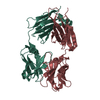
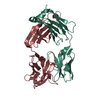
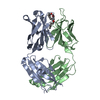
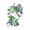
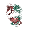
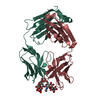
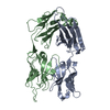
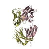
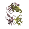
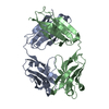
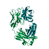
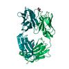
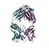
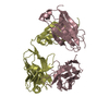
| Title | Structure of immature VRC01-class antibody DRVIA7 | ||||||
 Components Components |
| ||||||
 Keywords Keywords | IMMUNE SYSTEM / VRC01 / HIV-1 / CD4 binding site / gp120 | ||||||
| Function / homology | Immunoglobulins / Immunoglobulin-like / Sandwich / Mainly Beta Function and homology information Function and homology information | ||||||
| Biological species |  Homo sapiens (human) Homo sapiens (human) | ||||||
| Method |  X-RAY DIFFRACTION / SYNCHROTRON / MOLECULAR REPLACEMENT / Resolution: 2.9 Å X-RAY DIFFRACTION / SYNCHROTRON / MOLECULAR REPLACEMENT / Resolution: 2.9 Å | ||||||
 Authors Authors | Kong, L. / Wilson, I.A. | ||||||
 Citation Citation | Journal: Immunity / Year: 2016 Title: Key gp120 Glycans Pose Roadblocks to the Rapid Development of VRC01-Class Antibodies in an HIV-1-Infected Chinese Donor. Authors: Leopold Kong / Bin Ju / Yajing Chen / Linling He / Li Ren / Jiandong Liu / Kunxue Hong / Bin Su / Zheng Wang / Gabriel Ozorowski / Xiaolin Ji / Yuanzi Hua / Yanli Chen / Marc C Deller / ...Authors: Leopold Kong / Bin Ju / Yajing Chen / Linling He / Li Ren / Jiandong Liu / Kunxue Hong / Bin Su / Zheng Wang / Gabriel Ozorowski / Xiaolin Ji / Yuanzi Hua / Yanli Chen / Marc C Deller / Yanling Hao / Yi Feng / Fernando Garces / Richard Wilson / Kaifan Dai / Sijy O'Dell / Krisha McKee / John R Mascola / Andrew B Ward / Richard T Wyatt / Yuxing Li / Ian A Wilson / Jiang Zhu / Yiming Shao /   Abstract: VRC01-class antibodies neutralize diverse HIV-1 strains by targeting the conserved CD4-binding site. Despite extensive investigations, crucial events in the early stage of VRC01 development remain ...VRC01-class antibodies neutralize diverse HIV-1 strains by targeting the conserved CD4-binding site. Despite extensive investigations, crucial events in the early stage of VRC01 development remain elusive. We demonstrated how VRC01-class antibodies emerged in a Chinese donor by antigen-specific single B cell sorting, structural and functional studies, and longitudinal antibody and virus repertoire analyses. A monoclonal antibody DRVIA7 with modest neutralizing breadth was isolated that displayed a subset of VRC01 signatures. X-ray and EM structures revealed a VRC01-like angle of approach, but less favorable interactions between the DRVIA7 light-chain CDR1 and the N terminus with N276 and V5 glycans of gp120. Although the DRVIA7 lineage was unable to acquire broad neutralization, longitudinal analysis revealed a repertoire-encoded VRC01 light-chain CDR3 signature and VRC01-like neutralizing heavy-chain precursors that rapidly matured within 2 years. Thus, light chain accommodation of the glycan shield should be taken into account in vaccine design targeting this conserved site of vulnerability. | ||||||
| History |
|
- Structure visualization
| Structure viewer | Molecule: MolmilJmol/JSmol |
|---|
- Downloads & links
-Download
| PDBx/mmCIF format | 5cd3.cif.gz | 327.2 KB | Display | PDBx/mmCIF format |
|---|---|---|---|---|
| PDB format | pdb5cd3.ent.gz | 267.4 KB | Display | PDB format |
| PDBx/mmJSON format | 5cd3.json.gz | Tree view | PDBx/mmJSON format | |
| Others |  Other downloads Other downloads |
-Validation report
| Arichive directory | https://data.pdbj.org/pub/pdb/validation_reports/cd/5cd3ftp://data.pdbj.org/pub/pdb/validation_reports/cd/5cd3 | HTTPS FTP |
|---|
-Related structure data


-Links
 PDBj
PDBj


- Assembly
| Deposited unit | 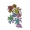
| ||||||||
|---|---|---|---|---|---|---|---|---|---|
| 1 | 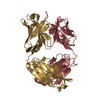
| ||||||||
| 2 | 
| ||||||||
| 3 | 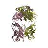
| ||||||||
| 4 | 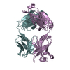
| ||||||||
| Unit cell |
|
-Components
| #1: Antibody | Mass: 24048.031 Da / Num. of mol.: 4 Source method: isolated from a genetically manipulated source Source: (gene. exp.) Homo sapiens (human) / Cell line (production host): HEK293F / Production host: Homo sapiens (human)#2: Antibody | Mass: 23207.871 Da / Num. of mol.: 4 Source method: isolated from a genetically manipulated source Source: (gene. exp.) Homo sapiens (human) / Cell line (production host): HEK293F / Production host: Homo sapiens (human)Has protein modification | Y | |
|---|
-Experimental details
-Experiment
| Experiment | Method: X-RAY DIFFRACTION / Number of used crystals: 1 |
|---|
- Sample preparation
| Crystal | Density Matthews: 2.64 Å3/Da / Density % sol: 53.48 % |
|---|---|
| Crystal grow | Temperature: 292 K / Method: vapor diffusion, sitting drop / pH: 4.6 Details: 0.16 M ammonium sulfate, 20% (w/v) PEG 4000, 20% (v/v) glycerol and 0.08 M sodium acetate, pH 4.6 |
-Data collection
| Diffraction | Mean temperature: 100 K |
|---|---|
| Diffraction source | Source: SYNCHROTRON / Site: APS / Beamline: 23-ID-B / Wavelength: 1.033 Å |
| Detector | Type: MAR CCD 130 mm / Detector: CCD / Date: Aug 9, 2014 |
| Radiation | Protocol: SINGLE WAVELENGTH / Monochromatic (M) / Laue (L): M / Scattering type: x-ray |
| Radiation wavelength | Wavelength: 1.033 Å / Relative weight: 1 |
| Reflection | Resolution: 2.9→50 Å / Num. obs: 41586 / % possible obs: 98.4 % / Redundancy: 1.8 % / Rmerge(I) obs: 0.24 / Net I/σ(I): 3.84 |
| Reflection shell | Resolution: 2.9→2.95 Å / Redundancy: 1.8 % / Rmerge(I) obs: 0.89 / Mean I/σ(I) obs: 1.1 / % possible all: 98.4 |
- Processing
| Software |
| ||||||||||||||||||||||||||||||||||||||||||||||||||||||||||||||||||||||||||||||||||||||||||||||||||||||||||||||||
|---|---|---|---|---|---|---|---|---|---|---|---|---|---|---|---|---|---|---|---|---|---|---|---|---|---|---|---|---|---|---|---|---|---|---|---|---|---|---|---|---|---|---|---|---|---|---|---|---|---|---|---|---|---|---|---|---|---|---|---|---|---|---|---|---|---|---|---|---|---|---|---|---|---|---|---|---|---|---|---|---|---|---|---|---|---|---|---|---|---|---|---|---|---|---|---|---|---|---|---|---|---|---|---|---|---|---|---|---|---|---|---|---|---|
| Refinement | Method to determine structure: MOLECULAR REPLACEMENT / Resolution: 2.9→49.542 Å / SU ML: 0.43 / Cross valid method: FREE R-VALUE / σ(F): 1.96 / Phase error: 30.66 / Stereochemistry target values: ML
| ||||||||||||||||||||||||||||||||||||||||||||||||||||||||||||||||||||||||||||||||||||||||||||||||||||||||||||||||
| Solvent computation | Shrinkage radii: 0.9 Å / VDW probe radii: 1.11 Å / Solvent model: FLAT BULK SOLVENT MODEL | ||||||||||||||||||||||||||||||||||||||||||||||||||||||||||||||||||||||||||||||||||||||||||||||||||||||||||||||||
| Refinement step | Cycle: LAST / Resolution: 2.9→49.542 Å
| ||||||||||||||||||||||||||||||||||||||||||||||||||||||||||||||||||||||||||||||||||||||||||||||||||||||||||||||||
| Refine LS restraints |
| ||||||||||||||||||||||||||||||||||||||||||||||||||||||||||||||||||||||||||||||||||||||||||||||||||||||||||||||||
| LS refinement shell |
|
