 Movie
Movie Controller

[English] 日本語
 Yorodumi
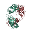
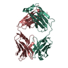
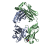
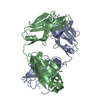
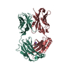
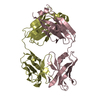
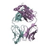
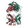
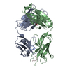
- PDB-3f12: Germline V-genes sculpt the binding site of a family of antibodie... -
+ Open data

- Basic information
| Entry | Database: PDB / ID: 3f12 | ||||||
|---|---|---|---|---|---|---|---|
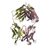
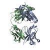
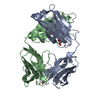
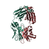
| Title | Germline V-genes sculpt the binding site of a family of antibodies neutralizing human cytomegalovirus | ||||||
 Components Components |
| ||||||
 Keywords Keywords | IMMUNE SYSTEM / Human cytomegalovirus / antibody / germline / Immunoglobulin domain | ||||||
| Function / homology | Immunoglobulins / Immunoglobulin-like / Sandwich / Mainly Beta Function and homology information Function and homology information | ||||||
| Biological species |  Homo sapiens (human) Homo sapiens (human) | ||||||
| Method |  X-RAY DIFFRACTION / SYNCHROTRON / MOLECULAR REPLACEMENT / Resolution: 2.95 Å X-RAY DIFFRACTION / SYNCHROTRON / MOLECULAR REPLACEMENT / Resolution: 2.95 Å | ||||||
 Authors Authors | Thomson, C.A. / Bryson, S. / McLean, G.R. / Creagh, A.L. / Pai, E.F. / Schrader, J.W. | ||||||
 Citation Citation | Journal: Embo J. / Year: 2008 Title: Germline V-genes sculpt the binding site of a family of antibodies neutralizing human cytomegalovirus. Authors: Thomson, C.A. / Bryson, S. / McLean, G.R. / Creagh, A.L. / Pai, E.F. / Schrader, J.W. | ||||||
| History |
|
- Structure visualization
| Structure viewer | Molecule: MolmilJmol/JSmol |
|---|
- Downloads & links
-Download
| PDBx/mmCIF format | 3f12.cif.gz | 166 KB | Display | PDBx/mmCIF format |
|---|---|---|---|---|
| PDB format | pdb3f12.ent.gz | 131.9 KB | Display | PDB format |
| PDBx/mmJSON format | 3f12.json.gz | Tree view | PDBx/mmJSON format | |
| Others |  Other downloads Other downloads |
-Validation report
| Arichive directory | https://data.pdbj.org/pub/pdb/validation_reports/f1/3f12ftp://data.pdbj.org/pub/pdb/validation_reports/f1/3f12 | HTTPS FTP |
|---|
-Related structure data
| Related structure data |  3eyfC  3eyoSC 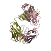 3eyqC C: citing same article ( S: Starting model for refinement |
|---|---|
| Similar structure data |


-Links
 PDBj
PDBj

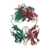
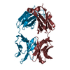
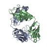
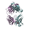
- Assembly
| Deposited unit | 
| ||||||||
|---|---|---|---|---|---|---|---|---|---|
| 1 | 
| ||||||||
| 2 | 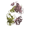
| ||||||||
| Unit cell |
|
-Components
| #1: Antibody | Mass: 23723.350 Da / Num. of mol.: 2 Source method: isolated from a genetically manipulated source Source: (gene. exp.) Homo sapiens (human) / Plasmid: pET32b / Production host:  #2: Antibody | Mass: 25842.934 Da / Num. of mol.: 2 Source method: isolated from a genetically manipulated source Source: (gene. exp.) Homo sapiens (human) / Plasmid: pET40 / Production host: Has protein modification | Y | |
|---|
-Experimental details
-Experiment
| Experiment | Method: X-RAY DIFFRACTION / Number of used crystals: 1 |
|---|
- Sample preparation
| Crystal | Density Matthews: 2.72 Å3/Da / Density % sol: 54.75 % |
|---|---|
| Crystal grow | Temperature: 298 K / Method: vapor diffusion, hanging drop / pH: 10.6 Details: 2.6 M Ammonium sulfate, 2% PEG 400 in CAPS buffer, pH 10.6, VAPOR DIFFUSION, HANGING DROP, temperature 298.0K |
-Data collection
| Diffraction | Mean temperature: 110 K |
|---|---|
| Diffraction source | Source: SYNCHROTRON / Site: APS  / Beamline: 14-BM-C / Wavelength: 0.9 Å / Beamline: 14-BM-C / Wavelength: 0.9 Å |
| Detector | Type: MARMOSAIC 225 mm CCD / Detector: CCD / Date: Mar 6, 2006 |
| Radiation | Monochromator: GRAPHITE / Protocol: SINGLE WAVELENGTH / Monochromatic (M) / Laue (L): M / Scattering type: x-ray |
| Radiation wavelength | Wavelength: 0.9 Å / Relative weight: 1 |
| Reflection | Resolution: 2.95→50 Å / Num. all: 23047 / Num. obs: 23047 / % possible obs: 98 % / Observed criterion σ(F): 0 / Observed criterion σ(I): 0 / Redundancy: 2.8 % / Rmerge(I) obs: 0.066 / Net I/σ(I): 19.12 |
| Reflection shell | Resolution: 2.95→3.1 Å / Redundancy: 2.7 % / Rmerge(I) obs: 0.333 / Mean I/σ(I) obs: 3.84 / % possible all: 98.4 |
- Processing
| Software |
| ||||||||||||||||||||
|---|---|---|---|---|---|---|---|---|---|---|---|---|---|---|---|---|---|---|---|---|---|
| Refinement | Method to determine structure: MOLECULAR REPLACEMENT Starting model: PDB entry 3EYO Resolution: 2.95→50 Å / Stereochemistry target values: Engh & Huber
| ||||||||||||||||||||
| Displacement parameters | Biso mean: 40.8 Å2 | ||||||||||||||||||||
| Refinement step | Cycle: LAST / Resolution: 2.95→50 Å
| ||||||||||||||||||||
| Refine LS restraints |
|
