 Movie
Movie Controller

[English] 日本語
 Yorodumi
- PDB-3eyf: Crystal structure of anti-human cytomegalovirus antibody 8f9 plus... -
+ Open data

- Basic information
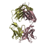
| Entry | Database: PDB / ID: 3eyf | ||||||
|---|---|---|---|---|---|---|---|
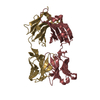
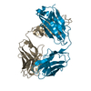
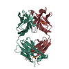
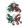
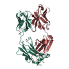
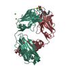
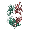
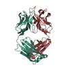
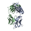
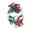
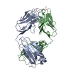
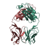
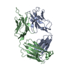
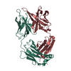
| Title | Crystal structure of anti-human cytomegalovirus antibody 8f9 plus gB peptide | ||||||
 Components Components |
| ||||||
 Keywords Keywords | IMMUNE SYSTEM / cytomegalovirus / antibody / Immunoglobulin domain / Cleavage on pair of basic residues / Envelope protein / Glycoprotein / Host-virus interaction / Membrane / Transmembrane / Virion | ||||||
| Function / homology |  Function and homology information Function and homology informationhost cell Golgi membrane / immunoglobulin complex / host cell endosome membrane / adaptive immune response / viral envelope / symbiont entry into host cell / virion attachment to host cell / host cell plasma membrane / virion membrane / extracellular region / plasma membrane Similarity search - Function | ||||||
| Biological species |  Homo sapiens (human) Homo sapiens (human) | ||||||
| Method |  X-RAY DIFFRACTION / MOLECULAR REPLACEMENT / Resolution: 2.3 Å X-RAY DIFFRACTION / MOLECULAR REPLACEMENT / Resolution: 2.3 Å | ||||||
 Authors Authors | Thomson, C.A. / Bryson, S. / McLean, G.R. / Creagh, A.L. / Pai, E.F. / Schrader, J.W. | ||||||
 Citation Citation | Journal: Embo J. / Year: 2008 Title: Germline V-genes sculpt the binding site of a family of antibodies neutralizing human cytomegalovirus. Authors: Thomson, C.A. / Bryson, S. / McLean, G.R. / Creagh, A.L. / Pai, E.F. / Schrader, J.W. | ||||||
| History |
|
- Structure visualization
| Structure viewer | Molecule: MolmilJmol/JSmol |
|---|
- Downloads & links
-Download
| PDBx/mmCIF format | 3eyf.cif.gz | 188.3 KB | Display | PDBx/mmCIF format |
|---|---|---|---|---|
| PDB format | pdb3eyf.ent.gz | 148.5 KB | Display | PDB format |
| PDBx/mmJSON format | 3eyf.json.gz | Tree view | PDBx/mmJSON format | |
| Others |  Other downloads Other downloads |
-Validation report
| Arichive directory | https://data.pdbj.org/pub/pdb/validation_reports/ey/3eyfftp://data.pdbj.org/pub/pdb/validation_reports/ey/3eyf | HTTPS FTP |
|---|
-Related structure data
| Related structure data |  3eyoC 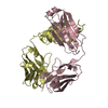 3eyqC 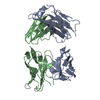 3f12C 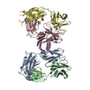 1hezS C: citing same article ( S: Starting model for refinement |
|---|---|
| Similar structure data |


-Links
 PDBj
PDBj

- Assembly
| Deposited unit | 
| ||||||||
|---|---|---|---|---|---|---|---|---|---|
| 1 | 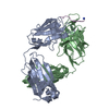
| ||||||||
| 2 | 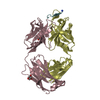
| ||||||||
| Unit cell |
|
-Components
| #1: Antibody | Mass: 23714.580 Da / Num. of mol.: 2 Source method: isolated from a genetically manipulated source Source: (gene. exp.) Homo sapiens (human) / Plasmid: pET32b / Production host:  #2: Antibody | Mass: 25932.000 Da / Num. of mol.: 2 Source method: isolated from a genetically manipulated source Source: (gene. exp.) Homo sapiens (human) / Plasmid: pET40 / Production host: #3: Protein/peptide | Mass: 1244.394 Da / Num. of mol.: 2 / Source method: obtained synthetically Details: Synthetic peptide following the sequence of residues 69-78 of Glycoprotein B from Human cytomegalovirus, UniProt entry P13201 (VGLB_HCMVT) References: UniProt: P13201 #4: Chemical |   Mass: 92.094 Da / Num. of mol.: 2 / Source method: obtained synthetically / Formula: C3H8O3 Mass: 92.094 Da / Num. of mol.: 2 / Source method: obtained synthetically / Formula: C3H8O3#5: Water | ChemComp-HOH / |  Mass: 18.015 Da / Num. of mol.: 402 / Source method: isolated from a natural source / Formula: H2O Mass: 18.015 Da / Num. of mol.: 402 / Source method: isolated from a natural source / Formula: H2OHas protein modification | Y | |
|---|
-Experimental details
-Experiment
| Experiment | Method: X-RAY DIFFRACTION / Number of used crystals: 1 |
|---|
- Sample preparation
| Crystal | Density Matthews: 2.45 Å3/Da / Density % sol: 49.86 % |
|---|---|
| Crystal grow | Temperature: 298 K / Method: vapor diffusion, hanging drop / pH: 10.6 Details: 1.9M Ammonium sulfate, 0.1M CAPS buffer, pH 10.6, VAPOR DIFFUSION, HANGING DROP, temperature 298K |
-Data collection
| Diffraction | Mean temperature: 110 K |
|---|---|
| Diffraction source | Source: ROTATING ANODE / Type: RIGAKU RU200 / Wavelength: 1.5418 Å |
| Detector | Type: RIGAKU RAXIS IV++ / Detector: IMAGE PLATE / Date: Sep 12, 2005 |
| Radiation | Monochromator: Ni filter / Protocol: SINGLE WAVELENGTH / Monochromatic (M) / Laue (L): M / Scattering type: x-ray |
| Radiation wavelength | Wavelength: 1.5418 Å / Relative weight: 1 |
| Reflection | Resolution: 2.3→50 Å / Num. all: 42862 / Num. obs: 42862 / % possible obs: 94.6 % / Observed criterion σ(F): 0 / Observed criterion σ(I): 0 / Redundancy: 3.1 % / Rmerge(I) obs: 0.051 / Net I/σ(I): 20.5 |
| Reflection shell | Resolution: 2.3→2.34 Å / Redundancy: 3.1 % / Rmerge(I) obs: 0.343 / Mean I/σ(I) obs: 4.5 / % possible all: 95.4 |
- Processing
| Software |
| ||||||||||||||||||||
|---|---|---|---|---|---|---|---|---|---|---|---|---|---|---|---|---|---|---|---|---|---|
| Refinement | Method to determine structure: MOLECULAR REPLACEMENT Starting model: PDB entry 1HEZ Resolution: 2.3→50 Å / σ(F): 0 / Stereochemistry target values: Engh & Huber
| ||||||||||||||||||||
| Displacement parameters | Biso mean: 36.8 Å2 | ||||||||||||||||||||
| Refinement step | Cycle: LAST / Resolution: 2.3→50 Å
| ||||||||||||||||||||
| Refine LS restraints |
|
