 Movie
Movie Controller


+ Open data

- Basic information
| Entry | Database: PDB / ID: 1hez | ||||||
|---|---|---|---|---|---|---|---|
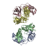
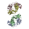
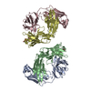
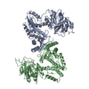
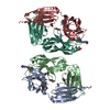
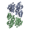
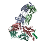
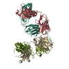
| Title | Structure of P. magnus protein L bound to a human IgM Fab. | ||||||
 Components Components |
| ||||||
 Keywords Keywords | ANTIBODY / SUPERANTIGEN | ||||||
| Function / homology |  Function and homology information Function and homology informationhexameric IgM immunoglobulin complex / IgM B cell receptor complex / pentameric IgM immunoglobulin complex / IgD immunoglobulin complex / IgA immunoglobulin complex / IgM immunoglobulin complex / IgE immunoglobulin complex / pre-B cell allelic exclusion / CD22 mediated BCR regulation / Fc epsilon receptor (FCERI) signaling ...hexameric IgM immunoglobulin complex / IgM B cell receptor complex / pentameric IgM immunoglobulin complex / IgD immunoglobulin complex / IgA immunoglobulin complex / IgM immunoglobulin complex / IgE immunoglobulin complex / pre-B cell allelic exclusion / CD22 mediated BCR regulation / Fc epsilon receptor (FCERI) signaling / IgG binding / Classical antibody-mediated complement activation / Initial triggering of complement / IgG immunoglobulin complex / immunoglobulin mediated immune response / FCGR activation / immunoglobulin binding / Role of LAT2/NTAL/LAB on calcium mobilization / Role of phospholipids in phagocytosis / Scavenging of heme from plasma / antigen binding / FCERI mediated Ca+2 mobilization / FCGR3A-mediated IL10 synthesis / Regulation of Complement cascade / Antigen activates B Cell Receptor (BCR) leading to generation of second messengers / Cell surface interactions at the vascular wall / B cell receptor signaling pathway / FCGR3A-mediated phagocytosis / FCERI mediated MAPK activation / Regulation of actin dynamics for phagocytic cup formation / FCERI mediated NF-kB activation / Immunoregulatory interactions between a Lymphoid and a non-Lymphoid cell / antibacterial humoral response / blood microparticle / Potential therapeutics for SARS / defense response to Gram-negative bacterium / adaptive immune response / immune response / innate immune response / cell surface / : / extracellular exosome / extracellular region / plasma membrane Similarity search - Function | ||||||
| Biological species |  HOMO SAPIENS (human) HOMO SAPIENS (human) FINEGOLDIA MAGNA (bacteria) FINEGOLDIA MAGNA (bacteria) | ||||||
| Method |  X-RAY DIFFRACTION / SYNCHROTRON / MOLECULAR REPLACEMENT / Resolution: 2.7 Å X-RAY DIFFRACTION / SYNCHROTRON / MOLECULAR REPLACEMENT / Resolution: 2.7 Å | ||||||
 Authors Authors | Graille, M. / Stura, E.A. / Housden, N.G. / Bottomley, S.P. / Taussig, M.J. / Sutton, B.J. / Gore, M.G. / Charbonnier, J.B. | ||||||
 Citation Citation | Journal: Structure / Year: 2001 Title: Complex between Peptostreptococcus Magnus Protein L and a Human Antibody Reveals Structural Convergence in the Interaction Modes of Fab Binding Modes Authors: Graille, M. / Stura, E.A. / Housden, N.G. / Beckingham, J.A. / Bottomley, S.P. / Beale, D. / Taussig, M.J. / Sutton, B.J. / Gore, M.G. / Charbonnier, J.B. #1: Journal: Biochem.J. / Year: 1999 Title: Interactions between a Single Immunoglobulin-Binding Domain of Protein L from Peptostreptococcus Magnus and a Human Kappa Light Chain Authors: Beckingham, J.A. / Bottomley, S.P. / Hinton, R. / Sutton, B.J. / Gore, M.G. #2: Journal: Proc.Natl.Acad.Sci.USA / Year: 2000Title: Crystal Structure of a Staphylococcus Aureus Protein a Domain Complexed with the Fab Fragment of a Human Igm Antibody: Structural Basis for Recognition of B-Cell Receptors and Superantigen Activity Authors: Graille, M. / Stura, E.A. / Corper, A.L. / Sutton, B.J. / Taussig, M.J. / Charbonnier, J.B. / Silverman, G.J. #3: Journal: Bioseparation / Year: 1995 Title: Cloning, expression and purification of Ppl-1, a kappa-chain binding protein, based upon protein L from Peptostreptococcus magnus. Authors: Bottomley, S.P. / Beckingham, J.A. / Murphy, J.P. / Atkinson, M. / Atkinson, T. / Hinton, R.J. / Gore, M.G. | ||||||
| History |
| ||||||
| Remark 700 | SHEET THE SHEET STRUCTURE OF THIS MOLECULE IS BIFURCATED. IN ORDER TO REPRESENT THIS FEATURE IN ... SHEET THE SHEET STRUCTURE OF THIS MOLECULE IS BIFURCATED. IN ORDER TO REPRESENT THIS FEATURE IN THE SHEET RECORDS BELOW, TWO SHEETS ARE DEFINED. |
- Structure visualization
| Structure viewer | Molecule: MolmilJmol/JSmol |
|---|
- Downloads & links
-Download
| PDBx/mmCIF format | 1hez.cif.gz | 183.8 KB | Display | PDBx/mmCIF format |
|---|---|---|---|---|
| PDB format | pdb1hez.ent.gz | 146.8 KB | Display | PDB format |
| PDBx/mmJSON format | 1hez.json.gz | Tree view | PDBx/mmJSON format | |
| Others |  Other downloads Other downloads |
-Validation report
| Arichive directory | https://data.pdbj.org/pub/pdb/validation_reports/he/1hezftp://data.pdbj.org/pub/pdb/validation_reports/he/1hez | HTTPS FTP |
|---|
-Related structure data
| Related structure data |  1deeS S: Starting model for refinement |
|---|---|
| Similar structure data |
-Links
 PDBj
PDBj


- Assembly
| Deposited unit | 
| ||||||||
|---|---|---|---|---|---|---|---|---|---|
| 1 |
| ||||||||
| Unit cell |
|
-Components
| #1: Antibody | Mass: 23373.871 Da / Num. of mol.: 2 / Fragment: 1-214 / Source method: isolated from a natural source / Source: (natural) HOMO SAPIENS (human) / Cell: B-LYMPHOCYTE / References: UniProt: P01834*PLUS#2: Antibody | Mass: 24292.139 Da / Num. of mol.: 2 / Fragment: 501-724 / Source method: isolated from a natural source / Source: (natural) HOMO SAPIENS (human) / Cell: B-LYMPHOCYTE / References: UniProt: P01871*PLUS#3: Protein | | Mass: 6750.485 Da / Num. of mol.: 1 / Fragment: 820-880 / Source method: isolated from a natural source / Source: (natural) FINEGOLDIA MAGNA (bacteria) / Plasmid: PKK223-3 / Strain: 3316 / References: UniProt: Q51918#4: Chemical |   Mass: 69.085 Da / Num. of mol.: 2 / Source method: obtained synthetically / Formula: C3H5N2 Mass: 69.085 Da / Num. of mol.: 2 / Source method: obtained synthetically / Formula: C3H5N2#5: Water | ChemComp-HOH / |  Mass: 18.015 Da / Num. of mol.: 112 / Source method: isolated from a natural source / Formula: H2O Mass: 18.015 Da / Num. of mol.: 112 / Source method: isolated from a natural source / Formula: H2OHas protein modification | Y | |
|---|
-Experimental details
-Experiment
| Experiment | Method: X-RAY DIFFRACTION / Number of used crystals: 1 |
|---|
- Sample preparation
| Crystal | Density Matthews: 2.13 Å3/Da / Density % sol: 63.1 % | ||||||||||||||||||||
|---|---|---|---|---|---|---|---|---|---|---|---|---|---|---|---|---|---|---|---|---|---|
| Crystal grow | pH: 8.5 Details: 13-16% MPEG 5000, 100MM IMIDAZOLE MALATE, PH8.5, pH 8.50 | ||||||||||||||||||||
| Crystal grow | *PLUS Method: vapor diffusion, sitting drop / Details: used seeding | ||||||||||||||||||||
| Components of the solutions | *PLUS
|
-Data collection
| Diffraction | Mean temperature: 120 K |
|---|---|
| Diffraction source | Source: SYNCHROTRON / Site: LURE  / Beamline: DW32 / Wavelength: 0.962 / Beamline: DW32 / Wavelength: 0.962 |
| Detector | Type: MAR scanner 345 mm plate / Detector: IMAGE PLATE / Date: Oct 15, 1999 |
| Radiation | Protocol: SINGLE WAVELENGTH / Monochromatic (M) / Laue (L): M / Scattering type: x-ray |
| Radiation wavelength | Wavelength: 0.962 Å / Relative weight: 1 |
| Reflection | Resolution: 2.7→20 Å / Num. obs: 26811 / % possible obs: 95.2 % / Redundancy: 4.66 % / Biso Wilson estimate: 59.53 Å2 / Rsym value: 0.112 / Net I/σ(I): 12.7 |
| Reflection shell | Resolution: 2.7→2.78 Å / Redundancy: 5 % / Mean I/σ(I) obs: 2.9 / Rsym value: 0.374 / % possible all: 97.6 |
| Reflection | *PLUS Highest resolution: 2.6 Å / Lowest resolution: 20 Å / Rmerge(I) obs: 0.116 |
| Reflection shell | *PLUS Highest resolution: 2.6 Å / Lowest resolution: 2.68 Å / % possible obs: 93.7 % / Rmerge(I) obs: 0.486 / Mean I/σ(I) obs: 2.5 |
- Processing
| Software |
| ||||||||||||||||||||||||||||||||||||||||||||||||||||||||||||
|---|---|---|---|---|---|---|---|---|---|---|---|---|---|---|---|---|---|---|---|---|---|---|---|---|---|---|---|---|---|---|---|---|---|---|---|---|---|---|---|---|---|---|---|---|---|---|---|---|---|---|---|---|---|---|---|---|---|---|---|---|---|
| Refinement | Method to determine structure: MOLECULAR REPLACEMENT Starting model: 1DEE Resolution: 2.7→20 Å / Rfactor Rfree error: 0.008 / Data cutoff high absF: 10000 / Cross valid method: THROUGHOUT / σ(F): 2 Details: SIDE CHAIN ATOMS FROM RESIDUES LYS A 45, LYS A 169, GLU B 89, LEU C 54, LYS C 107, GLU D 89, GLN D 113, GLU D 136, ARG D 181, LYS E 833 AND LYS E 878 ARE NOT DEFINED BY ELECTRONIC DENSITY
| ||||||||||||||||||||||||||||||||||||||||||||||||||||||||||||
| Solvent computation | Bsol: 22.3753 Å2 / ksol: 0.293557 e/Å3 | ||||||||||||||||||||||||||||||||||||||||||||||||||||||||||||
| Displacement parameters | Biso mean: 45.5 Å2 | ||||||||||||||||||||||||||||||||||||||||||||||||||||||||||||
| Refine analyze | Luzzati coordinate error obs: 0.34 Å / Luzzati d res low obs: 6 Å | ||||||||||||||||||||||||||||||||||||||||||||||||||||||||||||
| Refinement step | Cycle: LAST / Resolution: 2.7→20 Å
| ||||||||||||||||||||||||||||||||||||||||||||||||||||||||||||
| Refine LS restraints |
| ||||||||||||||||||||||||||||||||||||||||||||||||||||||||||||
| LS refinement shell | Resolution: 2.7→2.74 Å / Total num. of bins used: 23
| ||||||||||||||||||||||||||||||||||||||||||||||||||||||||||||
| Xplor file |
| ||||||||||||||||||||||||||||||||||||||||||||||||||||||||||||
| Software | *PLUS Name: CNS / Version: 1 / Classification: refinement | ||||||||||||||||||||||||||||||||||||||||||||||||||||||||||||
| LS refinement shell | *PLUS Lowest resolution: 2.75 Å |
