 Movie
Movie Controller


+ Open data

- Basic information
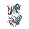
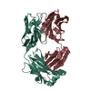
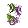
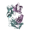
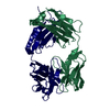
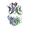
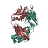
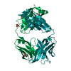
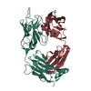
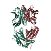
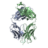
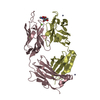
| Entry | Database: PDB / ID: 1dee | ||||||
|---|---|---|---|---|---|---|---|
| Title | Structure of S. aureus protein A bound to a human IgM Fab | ||||||
 Components Components |
| ||||||
 Keywords Keywords | IMMUNE SYSTEM / FAB-IBP COMPLEX CRYSTAL STRUCTURE 2.7A RESOLUTION BINDING OUTSIDE THE ANTIGEN COMBINING SITE SUPERANTIGEN FAB VH3 SPECIFICITY | ||||||
| Function / homology |  Function and homology information Function and homology informationsymbiont-mediated suppression of host signal transduction pathway via antagonism of host cell surface receptor / IgG binding / extracellular region Similarity search - Function | ||||||
| Biological species |   Staphylococcus aureus (bacteria) Staphylococcus aureus (bacteria) Homo sapiens (human) Homo sapiens (human) | ||||||
| Method |  X-RAY DIFFRACTION / Resolution: 2.7 Å X-RAY DIFFRACTION / Resolution: 2.7 Å | ||||||
 Authors Authors | Graille, M. / Stura, E.A. / Corper, A.L. / Sutton, B.J. / Taussig, M.J. / Charbonnier, J.B. / Silverman, G.J. | ||||||
 Citation Citation | Journal: Proc.Natl.Acad.Sci.USA / Year: 2000 Title: Crystal structure of a Staphylococcus aureus protein A domain complexed with the Fab fragment of a human IgM antibody: structural basis for recognition of B-cell receptors and superantigen activity. Authors: Graille, M. / Stura, E.A. / Corper, A.L. / Sutton, B.J. / Taussig, M.J. / Charbonnier, J.B. / Silverman, G.J. | ||||||
| History |
|
- Structure visualization
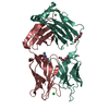
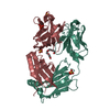
| Structure viewer | Molecule: MolmilJmol/JSmol |
|---|
- Downloads & links
-Download
| PDBx/mmCIF format | 1dee.cif.gz | 277.7 KB | Display | PDBx/mmCIF format |
|---|---|---|---|---|
| PDB format | pdb1dee.ent.gz | 222.6 KB | Display | PDB format |
| PDBx/mmJSON format | 1dee.json.gz | Tree view | PDBx/mmJSON format | |
| Others |  Other downloads Other downloads |
-Validation report
| Arichive directory | https://data.pdbj.org/pub/pdb/validation_reports/de/1deeftp://data.pdbj.org/pub/pdb/validation_reports/de/1dee | HTTPS FTP |
|---|
-Related structure data
| Similar structure data |
|---|


-Links
 PDBj
PDBj


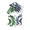
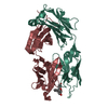
- Assembly
| Deposited unit | 
| ||||||||
|---|---|---|---|---|---|---|---|---|---|
| 1 | 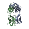
| ||||||||
| 2 | 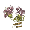
| ||||||||
| 3 | 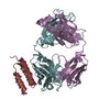
| ||||||||
| 4 | 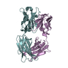
| ||||||||
| 5 | 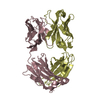
| ||||||||
| Unit cell |
|
-Components
| #1: Antibody | Mass: 23343.846 Da / Num. of mol.: 3 / Fragment: FAB LIGHT CHAIN / Source method: isolated from a natural source Details: FAB OF THE V3-30/ VH1.9III-ENCODED 2A2 IGM RHEUMATOID FACTOR PRODUCED BY TRYPSIN CLEAVAGE OF THE IGM SECRETED BY A HYBRIDOMA CREATED FROM SYNOVIAL B CELLS OF A RHEUMATOID ARTHRITIS PATIENT Source: (natural) Homo sapiens (human)#2: Antibody | Mass: 24167.008 Da / Num. of mol.: 3 / Fragment: FAB HEAVY CHAIN / Source method: isolated from a natural source Details: FAB OF THE V3-30/ VH1.9III-ENCODED 2A2 IGM RHEUMATOID FACTOR PRODUCED BY TRYPSIN CLEAVAGE OF THE IGM SECRETED BY A HYBRIDOMA CREATED FROM SYNOVIAL B CELLS OF A RHEUMATOID ARTHRITIS PATIENT Source: (natural) Homo sapiens (human)#3: Antibody | Mass: 6103.695 Da / Num. of mol.: 2 / Fragment: RECOMBINANT DOMAIN D Source method: isolated from a genetically manipulated source Source: (gene. exp.) Staphylococcus aureus (bacteria) / Production host: Staphylococcus aureus (bacteria) / References: UniProt: P02976#4: Water | ChemComp-HOH / |  Mass: 18.015 Da / Num. of mol.: 10 / Source method: isolated from a natural source / Formula: H2O Mass: 18.015 Da / Num. of mol.: 10 / Source method: isolated from a natural source / Formula: H2OHas protein modification | Y | |
|---|
-Experimental details
-Experiment
| Experiment | Method: X-RAY DIFFRACTION / Number of used crystals: 2 |
|---|
- Sample preparation
| Crystal | Density Matthews: 2.8 Å3/Da / Density % sol: 56.09 % | ||||||||||||||||||||
|---|---|---|---|---|---|---|---|---|---|---|---|---|---|---|---|---|---|---|---|---|---|
| Crystal grow | Temperature: 293 K / Method: vapor diffusion, sitting drop / pH: 6.5 Details: MPEG 5000, pH 6.5, VAPOR DIFFUSION, SITTING DROP, temperature 293K | ||||||||||||||||||||
| Crystal grow | *PLUS | ||||||||||||||||||||
| Components of the solutions | *PLUS
|
-Data collection
| Diffraction |
| |||||||||||||||
|---|---|---|---|---|---|---|---|---|---|---|---|---|---|---|---|---|
| Diffraction source |
| |||||||||||||||
| Detector |
| |||||||||||||||
| Radiation |
| |||||||||||||||
| Radiation wavelength | Wavelength: 1.5418 Å / Relative weight: 1 | |||||||||||||||
| Reflection | Resolution: 2.7→20 Å / Num. all: 184708 / Num. obs: 46831 / Biso Wilson estimate: 55 Å2 / Rmerge(I) obs: 0.065 | |||||||||||||||
| Reflection shell | Resolution: 2.7→2.8 Å / Rmerge(I) obs: 0.37 / % possible all: 77 | |||||||||||||||
| Reflection | *PLUS % possible obs: 87 % | |||||||||||||||
| Reflection shell | *PLUS % possible obs: 77 % / Mean I/σ(I) obs: 1.9 |
- Processing
| Software |
| ||||||||||||||||||||
|---|---|---|---|---|---|---|---|---|---|---|---|---|---|---|---|---|---|---|---|---|---|
| Refinement | Resolution: 2.7→10 Å / σ(F): 2 / σ(I): 0 / Stereochemistry target values: ENGH & HUBER Details: The 3 amino acids at the N terminus (FNK) of chain G have been removed for the refinement as they did not fit in any electronic density. However, the same amino acids have been kept in chain ...Details: The 3 amino acids at the N terminus (FNK) of chain G have been removed for the refinement as they did not fit in any electronic density. However, the same amino acids have been kept in chain H for refinement as more electron density is observed. The side chain atoms of Lys 805 from chain H are not defined by electronic density, then we decided to remove these atoms.
| ||||||||||||||||||||
| Refinement step | Cycle: LAST / Resolution: 2.7→10 Å
| ||||||||||||||||||||
| Software | *PLUS Name: X-PLOR / Version: 3.1 / Classification: refinement | ||||||||||||||||||||
| Refinement | *PLUS | ||||||||||||||||||||
| Solvent computation | *PLUS | ||||||||||||||||||||
| Displacement parameters | *PLUS | ||||||||||||||||||||
| Refine LS restraints | *PLUS
|
