 Movie
Movie Controller


+ Open data

- Basic information
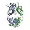
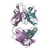
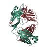
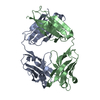
| Entry | Database: PDB / ID: 4ncc | ||||||
|---|---|---|---|---|---|---|---|
| Title | Neutralizing antibody to murine norovirus | ||||||
 Components Components |
| ||||||
 Keywords Keywords | IMMUNE SYSTEM / Immunoglobin / antibody / murine norovirus | ||||||
| Function / homology | Immunoglobulins / Immunoglobulin-like / Sandwich / Mainly Beta Function and homology information Function and homology information | ||||||
| Biological species |  | ||||||
| Method |  X-RAY DIFFRACTION / MOLECULAR REPLACEMENT / Resolution: 2.49 Å X-RAY DIFFRACTION / MOLECULAR REPLACEMENT / Resolution: 2.49 Å | ||||||
 Authors Authors | Smith, T. / Li, M. | ||||||
 Citation Citation | Journal: J.Virol. / Year: 2014 Title: Flexibility in surface-exposed loops in a virus capsid mediates escape from antibody neutralization. Authors: Kolawole, A.O. / Li, M. / Xia, C. / Fischer, A.E. / Giacobbi, N.S. / Rippinger, C.M. / Proescher, J.B. / Wu, S.K. / Bessling, S.L. / Gamez, M. / Yu, C. / Zhang, R. / Mehoke, T.S. / Pipas, J. ...Authors: Kolawole, A.O. / Li, M. / Xia, C. / Fischer, A.E. / Giacobbi, N.S. / Rippinger, C.M. / Proescher, J.B. / Wu, S.K. / Bessling, S.L. / Gamez, M. / Yu, C. / Zhang, R. / Mehoke, T.S. / Pipas, J.M. / Wolfe, J.T. / Lin, J.S. / Feldman, A.B. / Smith, T.J. / Wobus, C.E. | ||||||
| History |
|
- Structure visualization
| Structure viewer | Molecule: MolmilJmol/JSmol |
|---|
- Downloads & links
-Download
| PDBx/mmCIF format | 4ncc.cif.gz | 180.1 KB | Display | PDBx/mmCIF format |
|---|---|---|---|---|
| PDB format | pdb4ncc.ent.gz | 143 KB | Display | PDB format |
| PDBx/mmJSON format | 4ncc.json.gz | Tree view | PDBx/mmJSON format | |
| Others |  Other downloads Other downloads |
-Validation report
| Arichive directory | https://data.pdbj.org/pub/pdb/validation_reports/nc/4nccftp://data.pdbj.org/pub/pdb/validation_reports/nc/4ncc | HTTPS FTP |
|---|
-Related structure data
| Similar structure data |
|---|
-Links
 PDBj
PDBj

- Assembly
| Deposited unit | 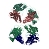
| |||||||||||||||||||||||||||||||||||||||||||||||||||||||||||
|---|---|---|---|---|---|---|---|---|---|---|---|---|---|---|---|---|---|---|---|---|---|---|---|---|---|---|---|---|---|---|---|---|---|---|---|---|---|---|---|---|---|---|---|---|---|---|---|---|---|---|---|---|---|---|---|---|---|---|---|---|
| 1 | 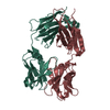
| |||||||||||||||||||||||||||||||||||||||||||||||||||||||||||
| 2 | 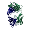
| |||||||||||||||||||||||||||||||||||||||||||||||||||||||||||
| Unit cell |
| |||||||||||||||||||||||||||||||||||||||||||||||||||||||||||
| Noncrystallographic symmetry (NCS) | NCS domain:
NCS domain segments:
NCS ensembles :
| |||||||||||||||||||||||||||||||||||||||||||||||||||||||||||
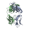
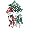
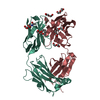
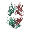
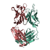
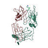
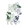
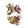
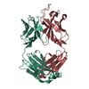
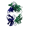
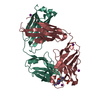
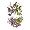
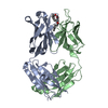
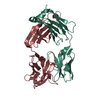
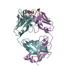
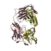
| Details | There are two Fabs in the asymmetric unit. L (light) and H (heavy) chains make one and chains 1 and 2 make the other where 1=light and 2=heavy |
-Components
| #1: Antibody | Mass: 23083.512 Da / Num. of mol.: 2 / Source method: isolated from a natural source / Source: (natural) #2: Antibody | Mass: 23759.061 Da / Num. of mol.: 2 / Source method: isolated from a natural source / Source: (natural) #3: Water | ChemComp-HOH / |  Mass: 18.015 Da / Num. of mol.: 370 / Source method: isolated from a natural source / Formula: H2O Mass: 18.015 Da / Num. of mol.: 370 / Source method: isolated from a natural source / Formula: H2OHas protein modification | Y | |
|---|
-Experimental details
-Experiment
| Experiment | Method: X-RAY DIFFRACTION / Number of used crystals: 1 |
|---|
- Sample preparation
| Crystal | Density Matthews: 2.57 Å3/Da / Density % sol: 52.06 % |
|---|---|
| Crystal grow | Temperature: 298 K / pH: 7.6 Details: Protein concentration 9.7 mg/ml in 50mM Tris. Reservoir 18% PEG 8000 in 90 mM Tris, pH 8.5. Drop was 5 microliters of reservoir and 5 of protein., VAPOR DIFFUSION, HANGING DROP, temperature 298K |
-Data collection
| Diffraction | Mean temperature: 100 K |
|---|---|
| Diffraction source | Source: ROTATING ANODE / Type: ENRAF-NONIUS FR591 / Wavelength: 1.5418 |
| Detector | Type: BRUKER SMART 6000 / Detector: CCD / Date: Jan 1, 2013 |
| Radiation | Monochromator: YALE MIRRORS / Protocol: SINGLE WAVELENGTH / Monochromatic (M) / Laue (L): M / Scattering type: x-ray |
| Radiation wavelength | Wavelength: 1.5418 Å / Relative weight: 1 |
| Reflection | Resolution: 2.49→42.49 Å / Num. obs: 20909 / % possible obs: 63 % / Observed criterion σ(I): 2 |
- Processing
| Software |
| |||||||||||||||||||||||||||||||||||||||||||||||||||||||||||||||
|---|---|---|---|---|---|---|---|---|---|---|---|---|---|---|---|---|---|---|---|---|---|---|---|---|---|---|---|---|---|---|---|---|---|---|---|---|---|---|---|---|---|---|---|---|---|---|---|---|---|---|---|---|---|---|---|---|---|---|---|---|---|---|---|---|
| Refinement | Method to determine structure: MOLECULAR REPLACEMENT / Resolution: 2.49→42.49 Å / Occupancy max: 1 / Occupancy min: 1 / SU ML: 0.34 / σ(F): 0 / Phase error: 26.79 / Stereochemistry target values: ML
| |||||||||||||||||||||||||||||||||||||||||||||||||||||||||||||||
| Solvent computation | Shrinkage radii: 0.9 Å / VDW probe radii: 1.11 Å / Solvent model: FLAT BULK SOLVENT MODEL | |||||||||||||||||||||||||||||||||||||||||||||||||||||||||||||||
| Displacement parameters | Biso mean: 18.96 Å2 | |||||||||||||||||||||||||||||||||||||||||||||||||||||||||||||||
| Refinement step | Cycle: LAST / Resolution: 2.49→42.49 Å
| |||||||||||||||||||||||||||||||||||||||||||||||||||||||||||||||
| Refine LS restraints |
| |||||||||||||||||||||||||||||||||||||||||||||||||||||||||||||||
| Refine LS restraints NCS |
| |||||||||||||||||||||||||||||||||||||||||||||||||||||||||||||||
| LS refinement shell |
|
