National Institutes of Health/National Institute Of Allergy and Infectious Diseases (NIH/NIAID)
R01 AI089591
United States
National Institutes of Health/National Institute Of Allergy and Infectious Diseases (NIH/NIAID)
R01 AI114816
United States
Citation
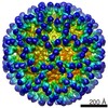
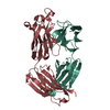
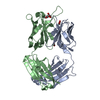
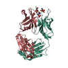
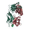
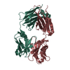
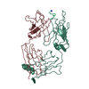
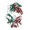
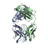
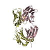
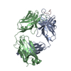
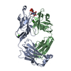
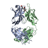
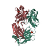
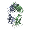
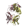
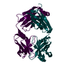
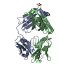
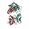
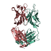
Journal: Proc Natl Acad Sci U S A / Year: 2015 Title: Cryo-EM structures elucidate neutralizing mechanisms of anti-chikungunya human monoclonal antibodies with therapeutic activity. Authors: Feng Long / Rachel H Fong / Stephen K Austin / Zhenguo Chen / Thomas Klose / Andrei Fokine / Yue Liu / Jason Porta / Gopal Sapparapu / Wataru Akahata / Benjamin J Doranz / James E Crowe / ...Authors: Feng Long / Rachel H Fong / Stephen K Austin / Zhenguo Chen / Thomas Klose / Andrei Fokine / Yue Liu / Jason Porta / Gopal Sapparapu / Wataru Akahata / Benjamin J Doranz / James E Crowe / Michael S Diamond / Michael G Rossmann / Abstract: Chikungunya virus (CHIKV) is a mosquito-transmitted alphavirus that causes severe acute and chronic disease in humans. Although highly inhibitory murine and human monoclonal antibodies (mAbs) have ...Chikungunya virus (CHIKV) is a mosquito-transmitted alphavirus that causes severe acute and chronic disease in humans. Although highly inhibitory murine and human monoclonal antibodies (mAbs) have been generated, the structural basis of their neutralizing activity remains poorly characterized. Here, we determined the cryo-EM structures of chikungunya virus-like particles complexed with antibody fragments (Fab) of two highly protective human mAbs, 4J21 and 5M16, that block virus fusion with host membranes. Both mAbs bind primarily to sites within the A and B domains, as well as to the B domain's β-ribbon connector of the viral glycoprotein E2. The footprints of these antibodies on the viral surface were consistent with results from loss-of-binding studies using an alanine scanning mutagenesis-based epitope mapping approach. The Fab fragments stabilized the position of the B domain relative to the virus, particularly for the complex with 5M16. This finding is consistent with a mechanism of neutralization in which anti-CHIKV mAbs that bridge the A and B domains impede movement of the B domain away from the underlying fusion loop on the E1 glycoprotein and therefore block the requisite pH-dependent fusion of viral and host membranes.
In the structure databanks used in Yorodumi, some data are registered as the other names, "COVID-19 virus" and "2019-nCoV". Here are the details of the virus and the list of structure data.
Jan 31, 2019. EMDB accession codes are about to change! (news from PDBe EMDB page)
EMDB accession codes are about to change! (news from PDBe EMDB page)
The allocation of 4 digits for EMDB accession codes will soon come to an end. Whilst these codes will remain in use, new EMDB accession codes will include an additional digit and will expand incrementally as the available range of codes is exhausted. The current 4-digit format prefixed with “EMD-” (i.e. EMD-XXXX) will advance to a 5-digit format (i.e. EMD-XXXXX), and so on. It is currently estimated that the 4-digit codes will be depleted around Spring 2019, at which point the 5-digit format will come into force.
The EM Navigator/Yorodumi systems omit the EMD- prefix.
Related info.:Q: What is EMD? / ID/Accession-code notation in Yorodumi/EM Navigator
Yorodumi is a browser for structure data from EMDB, PDB, SASBDB, etc.
This page is also the successor to EM Navigator detail page, and also detail information page/front-end page for Omokage search.
The word "yorodu" (or yorozu) is an old Japanese word meaning "ten thousand". "mi" (miru) is to see.
Related info.:EMDB / PDB / SASBDB / Comparison of 3 databanks / Yorodumi Search / Aug 31, 2016. New EM Navigator & Yorodumi / Yorodumi Papers / Jmol/JSmol / Function and homology information / Changes in new EM Navigator and Yorodumi
 Movie
Movie Controller

 Yorodumi
 Open data
 Basic information
 Components
 Keywords
 Function and homology information
 Homo sapiens (human)
 X-RAY DIFFRACTION /
  Authors
 United States, 2items
  Citation
 Structure visualization
 Downloads & links
 Other downloads
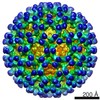


 PDBj
PDBj

 Assembly
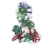

 Mass: 18.015 Da / Num. of mol.: 640 / Source method: isolated from a natural source / Formula: H2O
 Sample preparation
 Processing