 Movie
Movie Controller

[English] 日本語
 Yorodumi
- PDB-3lex: 2F5 Epitope scaffold elicited anti-HIV-1 monoclonal antibody 11F1... -
+ Open data

- Basic information
| Entry | Database: PDB / ID: 3lex | ||||||
|---|---|---|---|---|---|---|---|
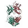
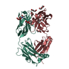
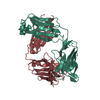
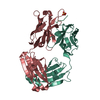
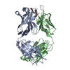
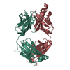
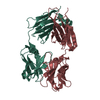
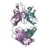
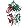
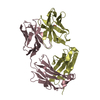
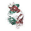
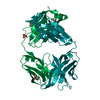
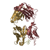
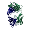
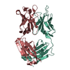
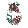
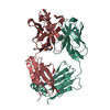
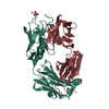
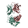
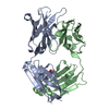
| Title | 2F5 Epitope scaffold elicited anti-HIV-1 monoclonal antibody 11F10 in complex with HIV-1 GP41 | ||||||
 Components Components |
| ||||||
 Keywords Keywords | IMMUNE SYSTEM / HIV-1 / GP-41 / MONOCLONAL ANTIBODY / 2F5 / SCAFFOLD / EPITOPE / TRANSPLANT / GRAFT / RE-ELCITATION / Envelope protein / VACCINE DESIGN | ||||||
| Function / homology |  Function and homology information Function and homology informationmembrane fusion involved in viral entry into host cell / viral envelope / symbiont entry into host cell / virion attachment to host cell / host cell plasma membrane / virion membrane / structural molecule activity Similarity search - Function | ||||||
| Biological species |    Human immunodeficiency virus 1 Human immunodeficiency virus 1 | ||||||
| Method |  X-RAY DIFFRACTION / SYNCHROTRON / MOLECULAR REPLACEMENT / Resolution: 1.97 Å X-RAY DIFFRACTION / SYNCHROTRON / MOLECULAR REPLACEMENT / Resolution: 1.97 Å | ||||||
 Authors Authors | Ofek, G. / Guenaga, F.J. / Schief, W.R. / Skinner, J. / Baker, D. / Wyatt, R. / Kwong, P.D. | ||||||
 Citation Citation | Journal: Proc.Natl.Acad.Sci.USA / Year: 2010 Title: Elicitation of structure-specific antibodies by epitope scaffolds. Authors: Ofek, G. / Guenaga, F.J. / Schief, W.R. / Skinner, J. / Baker, D. / Wyatt, R. / Kwong, P.D. | ||||||
| History |
|
- Structure visualization
| Structure viewer | Molecule: MolmilJmol/JSmol |
|---|
- Downloads & links
-Download
| PDBx/mmCIF format | 3lex.cif.gz | 192.1 KB | Display | PDBx/mmCIF format |
|---|---|---|---|---|
| PDB format | pdb3lex.ent.gz | 151.4 KB | Display | PDB format |
| PDBx/mmJSON format | 3lex.json.gz | Tree view | PDBx/mmJSON format | |
| Others |  Other downloads Other downloads |
-Validation report
| Arichive directory | https://data.pdbj.org/pub/pdb/validation_reports/le/3lexftp://data.pdbj.org/pub/pdb/validation_reports/le/3lex | HTTPS FTP |
|---|
-Related structure data
| Related structure data | 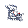 3lesC 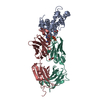 3levC 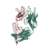 3leySC C: citing same article ( S: Starting model for refinement |
|---|---|
| Similar structure data |

-Links
 PDBj
PDBj


- Assembly
| Deposited unit | 
| ||||||||
|---|---|---|---|---|---|---|---|---|---|
| 1 | 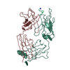
| ||||||||
| 2 | 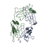
| ||||||||
| Unit cell |
|
-Components
| #1: Antibody | Mass: 23916.674 Da / Num. of mol.: 2 / Source method: isolated from a natural source / Source: (natural) #2: Antibody | Mass: 24264.043 Da / Num. of mol.: 2 / Source method: isolated from a natural source / Source: (natural) #3: Protein/peptide | Mass: 986.165 Da / Num. of mol.: 2 / Source method: obtained synthetically / Details: HIV-1 gp41 peptide / Source: (synth.) Human immunodeficiency virus 1 / References: UniProt: Q9IJQ0#4: Water | ChemComp-HOH / |  Mass: 18.015 Da / Num. of mol.: 628 / Source method: isolated from a natural source / Formula: H2O Mass: 18.015 Da / Num. of mol.: 628 / Source method: isolated from a natural source / Formula: H2OHas protein modification | Y | |
|---|
-Experimental details
-Experiment
| Experiment | Method: X-RAY DIFFRACTION / Number of used crystals: 1 |
|---|
- Sample preparation
| Crystal | Density Matthews: 1.97 Å3/Da / Density % sol: 37.44 % |
|---|---|
| Crystal grow | Temperature: 293 K / Method: vapor diffusion, hanging drop Details: 16% PEG 4000, 0.08 M CH3COONA, 0.04 M, TRIS PH 8.5, VAPOR DIFFUSION, HANGING DROP, temperature 293K |
-Data collection
| Diffraction | Mean temperature: 100 K |
|---|---|
| Diffraction source | Source: SYNCHROTRON / Site: APS  / Beamline: 22-BM / Wavelength: 1 Å / Beamline: 22-BM / Wavelength: 1 Å |
| Detector | Type: MARMOSAIC 300 mm CCD / Detector: CCD / Date: Apr 2, 2009 |
| Radiation | Monochromator: SI (220) / Protocol: SINGLE WAVELENGTH / Monochromatic (M) / Laue (L): M / Scattering type: x-ray |
| Radiation wavelength | Wavelength: 1 Å / Relative weight: 1 |
| Reflection | Resolution: 1.96→25.9 Å / Num. obs: 49925 / % possible obs: 94.6 % / Observed criterion σ(I): 5 / Biso Wilson estimate: 19.46 Å2 / Rsym value: 0.038 / Net I/σ(I): 33 |
| Reflection shell | Resolution: 1.97→2.04 Å / Mean I/σ(I) obs: 12.9 / Rsym value: 0.092 / % possible all: 94.7 |
- Processing
| Software |
| |||||||||||||||||||||||||||||||||||||||||||||||||||||||||||||||||||||||||||||||||||||||||||||||||||||||||
|---|---|---|---|---|---|---|---|---|---|---|---|---|---|---|---|---|---|---|---|---|---|---|---|---|---|---|---|---|---|---|---|---|---|---|---|---|---|---|---|---|---|---|---|---|---|---|---|---|---|---|---|---|---|---|---|---|---|---|---|---|---|---|---|---|---|---|---|---|---|---|---|---|---|---|---|---|---|---|---|---|---|---|---|---|---|---|---|---|---|---|---|---|---|---|---|---|---|---|---|---|---|---|---|---|---|---|
| Refinement | Method to determine structure: MOLECULAR REPLACEMENT Starting model: 3LEY Resolution: 1.97→25.9 Å / Occupancy max: 1 / Occupancy min: 1 / SU ML: 1.16 / σ(F): 0.06 / Stereochemistry target values: ML
| |||||||||||||||||||||||||||||||||||||||||||||||||||||||||||||||||||||||||||||||||||||||||||||||||||||||||
| Solvent computation | Shrinkage radii: 0.9 Å / VDW probe radii: 1.11 Å / Solvent model: FLAT BULK SOLVENT MODEL / Bsol: 50.85 Å2 / ksol: 0.34 e/Å3 | |||||||||||||||||||||||||||||||||||||||||||||||||||||||||||||||||||||||||||||||||||||||||||||||||||||||||
| Displacement parameters | Biso mean: 26.95 Å2
| |||||||||||||||||||||||||||||||||||||||||||||||||||||||||||||||||||||||||||||||||||||||||||||||||||||||||
| Refinement step | Cycle: LAST / Resolution: 1.97→25.9 Å
| |||||||||||||||||||||||||||||||||||||||||||||||||||||||||||||||||||||||||||||||||||||||||||||||||||||||||
| Refine LS restraints |
| |||||||||||||||||||||||||||||||||||||||||||||||||||||||||||||||||||||||||||||||||||||||||||||||||||||||||
| LS refinement shell |
|
