- PDB-5dlm: Complex of Influenza M2e and Antibody -
+
Open data
ID or keywords:
Loading...
-
Basic information
Entry
Database: PDB / ID: 5dlm
Title
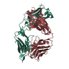
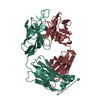
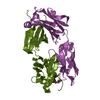
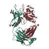
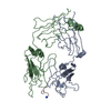
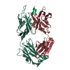
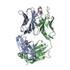
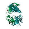
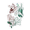
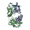
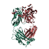
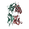
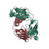
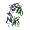
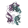
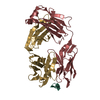
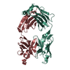
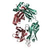
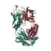
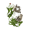
Complex of Influenza M2e and Antibody
Components
Heavy chain of monoclonal antibody
Light chain of monoclonal antibody
Matrix protein 2
Keywords
IMMUNE SYSTEM / Complex / Antibody / Extracellular domain
Function / homology
Function and homology information
uncoating of virus / : / Transport of HA trimer, NA tetramer and M2 tetramer from the endoplasmic reticulum to the Golgi Apparatus / Assembly of Viral Components at the Budding Site / Influenza Infection / Fusion of the Influenza Virion to the Host Cell Endosome / Release / Budding / Packaging of Eight RNA Segments / Uncoating of the Influenza Virion ...uncoating of virus / : / Transport of HA trimer, NA tetramer and M2 tetramer from the endoplasmic reticulum to the Golgi Apparatus / Assembly of Viral Components at the Budding Site / Influenza Infection / Fusion of the Influenza Virion to the Host Cell Endosome / Release / Budding / Packaging of Eight RNA Segments / Uncoating of the Influenza Virion / Entry of Influenza Virion into Host Cell via Endocytosis / proton transmembrane transporter activity / protein complex oligomerization / monoatomic ion channel activity / Viral mRNA Translation / symbiont genome entry into host cell via pore formation in plasma membrane / host cell plasma membrane / virion membrane / extracellular region / membrane / plasma membrane Similarity search - Function
H: Heavy chain of monoclonal antibody L: Light chain of monoclonal antibody I: Heavy chain of monoclonal antibody M: Light chain of monoclonal antibody X: Matrix protein 2 Y: Matrix protein 2 hetero molecules
In the structure databanks used in Yorodumi, some data are registered as the other names, "COVID-19 virus" and "2019-nCoV". Here are the details of the virus and the list of structure data.
Jan 31, 2019. EMDB accession codes are about to change! (news from PDBe EMDB page)
EMDB accession codes are about to change! (news from PDBe EMDB page)
The allocation of 4 digits for EMDB accession codes will soon come to an end. Whilst these codes will remain in use, new EMDB accession codes will include an additional digit and will expand incrementally as the available range of codes is exhausted. The current 4-digit format prefixed with “EMD-” (i.e. EMD-XXXX) will advance to a 5-digit format (i.e. EMD-XXXXX), and so on. It is currently estimated that the 4-digit codes will be depleted around Spring 2019, at which point the 5-digit format will come into force.
The EM Navigator/Yorodumi systems omit the EMD- prefix.
Related info.:Q: What is EMD? / ID/Accession-code notation in Yorodumi/EM Navigator
Yorodumi is a browser for structure data from EMDB, PDB, SASBDB, etc.
This page is also the successor to EM Navigator detail page, and also detail information page/front-end page for Omokage search.
The word "yorodu" (or yorozu) is an old Japanese word meaning "ten thousand". "mi" (miru) is to see.
Related info.:EMDB / PDB / SASBDB / Comparison of 3 databanks / Yorodumi Search / Aug 31, 2016. New EM Navigator & Yorodumi / Yorodumi Papers / Jmol/JSmol / Function and homology information / Changes in new EM Navigator and Yorodumi
 Movie
Movie Controller


 Open data
 Basic information
 Components
 Keywords
 Function and homology information

 Influenza A virus
 X-RAY DIFFRACTION /
  Authors
 Belgium,
  China, 5items
  Citation
 Structure visualization
 Downloads & links
 Other downloads


 PDBj
PDBj


 Assembly
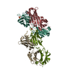

 Mass: 96.063 Da / Num. of mol.: 2 / Source method: obtained synthetically / Formula: SO4
 Mass: 18.015 Da / Num. of mol.: 305 / Source method: isolated from a natural source / Formula: H2O
 Sample preparation
 / Beamline: I24 / Wavelength: 1 Å
 Processing